Mystery of Bruch’s membrane explored
Wide-ranging Kreissig Lecture discussed theories on myopic axial elongation

Dermot McGrath
Published: Saturday, September 7, 2019
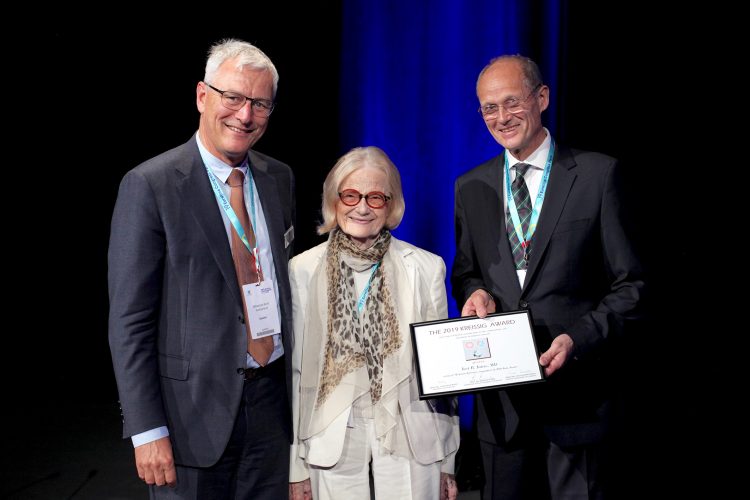 EURETINA President Sebastian Wolf (left) and Ingrid Kreissig present the Ingrid Kreissig Award to Jost Jonas
BRUCH’S membrane may play a far more significant role in the complex process of emmetropisation and myopisation of the human eye than it is currently given credit for, Jost Jonas MD said in the Kreissig Lecture delivered at the 19th EURETINA Congress.
“The more we learn about Bruch's membrane (BM), the more we realise that it may be more than just an almost invisible double basal membrane with some collagen and elastin in between,” he said.
In a wide-ranging lecture on “Myopia: Epidemiology, histology and clinics”, Prof Jonas, Professor of Ophthalmology of the Medical Faculty Mannheim of the Ruprecht-Karls-University Heidelberg, Germany, discussed his theory that myopic axial elongation of the globe may be influenced by an enlargement of Bruch’s membrane in the equatorial and retro-equatorial region of the eye.
BM is a five-layered inner limiting membrane of the retina separating the pigmented layer of the retina from the choroid of the eye, with the retinal pigment epithelium transporting metabolic waste from the photoreceptors across BM to the choroid.
Prof Jonas explained how the process of emmetropisation occurs by axial elongation soon after birth.
“Up to the end of the second year of life, the eye grows spherically by an active increase in scleral volume. Beyond that age, the process of emmetropisation begins in earnest with fine-tuning of the optical axis length to the optical characteristics of the lens and cornea. The axial elongation in the process of emmetropisation is associated with thinning of the retina and reduced density of retinal pigment epithelium cells (RPE) in the retro-equatorial region,” he said.
This thinning affects the choroid more than the sclera, starting at the equator and being most marked at the posterior pole, Dr Jonas added. By contrast, retinal thickness and RPE density in the macular region and thickness of BM in any region are independent of axial length.
The increased disc-fovea distance in axially myopic eyes is caused by the development and enlargement of parapapillary, BM-free gamma zone, while the length of macular BM, and indirectly RPE cell density and retinal thickness, remain constant.
Prof Jonas said that if BM is indeed the primary driver of axial elongation, leading to choroidal compression, then RPE may be the ideal target tissue for modification of emmetropisation and myopisation, producing and elongating BM in the retro-equatorial region.
A full account of Dr Jonas’s lecture will appear in a future edition of EuroTimes
EURETINA President Sebastian Wolf (left) and Ingrid Kreissig present the Ingrid Kreissig Award to Jost Jonas
BRUCH’S membrane may play a far more significant role in the complex process of emmetropisation and myopisation of the human eye than it is currently given credit for, Jost Jonas MD said in the Kreissig Lecture delivered at the 19th EURETINA Congress.
“The more we learn about Bruch's membrane (BM), the more we realise that it may be more than just an almost invisible double basal membrane with some collagen and elastin in between,” he said.
In a wide-ranging lecture on “Myopia: Epidemiology, histology and clinics”, Prof Jonas, Professor of Ophthalmology of the Medical Faculty Mannheim of the Ruprecht-Karls-University Heidelberg, Germany, discussed his theory that myopic axial elongation of the globe may be influenced by an enlargement of Bruch’s membrane in the equatorial and retro-equatorial region of the eye.
BM is a five-layered inner limiting membrane of the retina separating the pigmented layer of the retina from the choroid of the eye, with the retinal pigment epithelium transporting metabolic waste from the photoreceptors across BM to the choroid.
Prof Jonas explained how the process of emmetropisation occurs by axial elongation soon after birth.
“Up to the end of the second year of life, the eye grows spherically by an active increase in scleral volume. Beyond that age, the process of emmetropisation begins in earnest with fine-tuning of the optical axis length to the optical characteristics of the lens and cornea. The axial elongation in the process of emmetropisation is associated with thinning of the retina and reduced density of retinal pigment epithelium cells (RPE) in the retro-equatorial region,” he said.
This thinning affects the choroid more than the sclera, starting at the equator and being most marked at the posterior pole, Dr Jonas added. By contrast, retinal thickness and RPE density in the macular region and thickness of BM in any region are independent of axial length.
The increased disc-fovea distance in axially myopic eyes is caused by the development and enlargement of parapapillary, BM-free gamma zone, while the length of macular BM, and indirectly RPE cell density and retinal thickness, remain constant.
Prof Jonas said that if BM is indeed the primary driver of axial elongation, leading to choroidal compression, then RPE may be the ideal target tissue for modification of emmetropisation and myopisation, producing and elongating BM in the retro-equatorial region.
A full account of Dr Jonas’s lecture will appear in a future edition of EuroTimes