Paediatric keratoplasty
Pearls and pitfalls of keratoplasty in children.

Dermot McGrath
Published: Friday, November 1, 2019
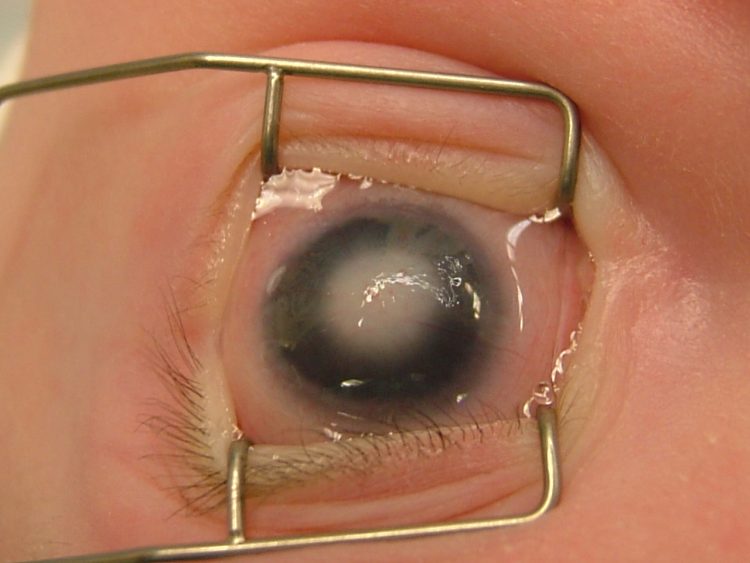 Preoperative image of a paediatric patient undergoing corneal transplantation. Courtesy of Gerald W. Zaidman MD, FAAO, FACS
Corneal transplantation in a paediatric patient population poses special challenges, but good results may still be obtained with careful case selection, rigorous surgery and meticulous postoperative care, according to Gerald W. Zaidman MD, FAAO, FACS.
Speaking during a World Society of Paediatric Ophthalmology and Strabismus (WSPOS) symposium held during the European Society of Ophthalmology (SOE) meeting in Nice, France, Dr Zaidman said that paediatric corneal diseases remain a frequent cause of blindness in the developed and developing world, accounting for an estimated 10% of all cases of blindness in children.
He noted that attempting keratoplasty in children is very different from adults and requires a fundamentally different approach.
“The challenge is that we are dealing with severe ocular pathology in a technically difficult surgical situation with a small eye, an elastic sclera and very shallow anterior chamber, with anterior displacement of the iris and lens, a young patient who does not cooperate, who is hard to examine and who can reject their corneas literally overnight,” he said.
The key first step is to accurately diagnose and manage a child with a cloudy cornea, said Dr Zaidman, who is Professor of Ophthalmology, New York Medical College, and Vice Chairman of Ophthalmology, Westchester Medical Center, both in Valhalla, New York, USA.
“There is every likelihood that you are going to have to perform an exam under anaesthesia (EUA) in a child under the age of three or four. You have to accurately diagnose and manage all the problems and conditions that may be causing the clouded cornea. This means performing all the exams, monitoring the IOP, checking the corneal diameter and performing A/B ultrasound scans. I would also advise most importantly to use ultrasound biomicroscopy (UBM) because that will help you to visualise any anterior segment changes,” he said.
The indications for paediatric keratoplasty are also markedly different than adult corneal transplant patients, the vast majority of whom have endothelial disease, keratoconus or failed grafts, said Dr Zaidman.
“I have performed over 400 transplants in children. Congenital diseases easily make up the vast bulk of the patients, primarily Peters’ anomaly, sclerocornea, endothelial dystrophy and congenital glaucoma. Trauma is the second single biggest indication and then in older children and adolescents we encounter keratoconus, herpes, corneal ulcers and rosacea,” he said.
A key part of the preoperative assessment entails an assessment of the social situation of the child and ensuring that the parents and caregivers are fully aware of what treatment will entail.
“They need to understand that this will be a marathon of eye drops, visits and examinations, many of them under general anaesthesia. We also need to realistically discuss success rates with them and explain that a clear cornea does not necessarily equate to good vision, and that treatment of amblyopia and glaucoma may severely impact on the outcomes,” he said.
In newborn patients with congenital corneal disease, Dr Zaidman advised scheduling the first office visit prior to three weeks of age and then performing the first exam under anaesthesia at four-to-six weeks.
Surgery in the first eye is usually performed between eight-and-12 weeks of age and in the second eye from four-to-six weeks after the first surgery. For the surgery, Dr Zaidman said that all patients undergo general anaesthesia, with mannitol and hyperventilation to ensure a softer globe and optimal conditions for the keratoplasty.
Donor tissue is used from donors aged four-to-19 years and the donor tissue is oversized by 0.5mm. A scleral support ring is then put in place and the anterior chamber is entered carefully using plenty of viscoelastic before synechialysis is performed, said Dr Zaidman.
Over the years, Dr Zaidman has developed what he calls the ‘sandwich technique’ in which the donor cornea is placed on to the host cornea and sutured into position into the recipient’s sclera.
Once this has been done, the recipient’s cornea is then removed from under the donor cornea, avoiding vitreous and lens prolapse. Interrupted sutures then are used to adhere the graft in place, Dr Zaidman explained.
“This technique works well and helps to minimise the extreme positive pressure typically encountered during paediatric surgery and to ensure a stable anterior chamber,” he said.
In terms of postoperative care, the patient is examined two-to-three times weekly and eye exams under anaesthesia are performed frequently. Sutures are removed within one month for infants and three-to-four months for young children. Optical correction is advised as soon as possible after suture removal.
Over the long term, topical steroids are tapered slowly over the course of one year and no vaccinations are administered for at least one year because of the risk of rejection.
In terms of results, Dr Zaidman said that patients with Peters’ anomaly type I usually obtained the best visual outcomes, with more than 85% having clear grafts and 54% obtaining visual acuity of 20/100 or better after three years. Children with glaucoma generally had the worst prognosis, he added.
Preoperative image of a paediatric patient undergoing corneal transplantation. Courtesy of Gerald W. Zaidman MD, FAAO, FACS
Corneal transplantation in a paediatric patient population poses special challenges, but good results may still be obtained with careful case selection, rigorous surgery and meticulous postoperative care, according to Gerald W. Zaidman MD, FAAO, FACS.
Speaking during a World Society of Paediatric Ophthalmology and Strabismus (WSPOS) symposium held during the European Society of Ophthalmology (SOE) meeting in Nice, France, Dr Zaidman said that paediatric corneal diseases remain a frequent cause of blindness in the developed and developing world, accounting for an estimated 10% of all cases of blindness in children.
He noted that attempting keratoplasty in children is very different from adults and requires a fundamentally different approach.
“The challenge is that we are dealing with severe ocular pathology in a technically difficult surgical situation with a small eye, an elastic sclera and very shallow anterior chamber, with anterior displacement of the iris and lens, a young patient who does not cooperate, who is hard to examine and who can reject their corneas literally overnight,” he said.
The key first step is to accurately diagnose and manage a child with a cloudy cornea, said Dr Zaidman, who is Professor of Ophthalmology, New York Medical College, and Vice Chairman of Ophthalmology, Westchester Medical Center, both in Valhalla, New York, USA.
“There is every likelihood that you are going to have to perform an exam under anaesthesia (EUA) in a child under the age of three or four. You have to accurately diagnose and manage all the problems and conditions that may be causing the clouded cornea. This means performing all the exams, monitoring the IOP, checking the corneal diameter and performing A/B ultrasound scans. I would also advise most importantly to use ultrasound biomicroscopy (UBM) because that will help you to visualise any anterior segment changes,” he said.
The indications for paediatric keratoplasty are also markedly different than adult corneal transplant patients, the vast majority of whom have endothelial disease, keratoconus or failed grafts, said Dr Zaidman.
“I have performed over 400 transplants in children. Congenital diseases easily make up the vast bulk of the patients, primarily Peters’ anomaly, sclerocornea, endothelial dystrophy and congenital glaucoma. Trauma is the second single biggest indication and then in older children and adolescents we encounter keratoconus, herpes, corneal ulcers and rosacea,” he said.
A key part of the preoperative assessment entails an assessment of the social situation of the child and ensuring that the parents and caregivers are fully aware of what treatment will entail.
“They need to understand that this will be a marathon of eye drops, visits and examinations, many of them under general anaesthesia. We also need to realistically discuss success rates with them and explain that a clear cornea does not necessarily equate to good vision, and that treatment of amblyopia and glaucoma may severely impact on the outcomes,” he said.
In newborn patients with congenital corneal disease, Dr Zaidman advised scheduling the first office visit prior to three weeks of age and then performing the first exam under anaesthesia at four-to-six weeks.
Surgery in the first eye is usually performed between eight-and-12 weeks of age and in the second eye from four-to-six weeks after the first surgery. For the surgery, Dr Zaidman said that all patients undergo general anaesthesia, with mannitol and hyperventilation to ensure a softer globe and optimal conditions for the keratoplasty.
Donor tissue is used from donors aged four-to-19 years and the donor tissue is oversized by 0.5mm. A scleral support ring is then put in place and the anterior chamber is entered carefully using plenty of viscoelastic before synechialysis is performed, said Dr Zaidman.
Over the years, Dr Zaidman has developed what he calls the ‘sandwich technique’ in which the donor cornea is placed on to the host cornea and sutured into position into the recipient’s sclera.
Once this has been done, the recipient’s cornea is then removed from under the donor cornea, avoiding vitreous and lens prolapse. Interrupted sutures then are used to adhere the graft in place, Dr Zaidman explained.
“This technique works well and helps to minimise the extreme positive pressure typically encountered during paediatric surgery and to ensure a stable anterior chamber,” he said.
In terms of postoperative care, the patient is examined two-to-three times weekly and eye exams under anaesthesia are performed frequently. Sutures are removed within one month for infants and three-to-four months for young children. Optical correction is advised as soon as possible after suture removal.
Over the long term, topical steroids are tapered slowly over the course of one year and no vaccinations are administered for at least one year because of the risk of rejection.
In terms of results, Dr Zaidman said that patients with Peters’ anomaly type I usually obtained the best visual outcomes, with more than 85% having clear grafts and 54% obtaining visual acuity of 20/100 or better after three years. Children with glaucoma generally had the worst prognosis, he added.
Tags: paediatric keratoplasty
Latest Articles
Making Female Leadership More than a Moment
A remarkable global confluence of women in key positions.
ESCRS Talks Technology at AAO
Europe adopts technological advances, US still waiting for lenses and lasers.
Sorting Out Simultaneous Vision IOLs
The ESCRS Eye Journal Club discuss a new landmark paper on IOL classification and the need for harmonisation of terminology for presbyopic IOLs.
Big Advantages to Small-Aperture IOLs
Small-aperture IOLs offer superior image quality with increased range of focus.
Prioritising Self-Care
Benefits of maintaining physical, emotional, and mental health extend beyond the personal sphere.
Valuing Clinical Trial Design
How inclusivity and diversity can enhance scientific accuracy in research.
Knowing Iris Repair: Using Iridodiathermy in Iris Surgery
Prepare for decentred pupils and uneven irides in multiple situations.
Neuroprotectant Treatment for MacTel Type 2
Intravitreal implant releasing ciliary neurotrophic factor found safe and effective in pivotal trials.