Cataract
Universalising Intraoperative OCT Benefits
Cost-effectiveness research needed to justify wider use of intraoperative OCT.
Howard Larkin
Published: Friday, December 1, 2023
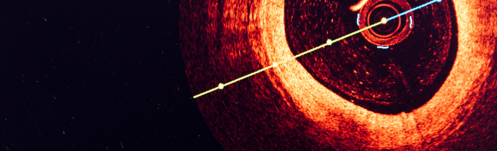
Optical coherence tomography (OCT) has revolutionised ophthalmic imaging, providing invaluable diagnostic information for both posterior and anterior segment structures. More recently, intraoperative OCT systems developed to help guide surgery are now integrated with operating microscopes.
But does intraoperative OCT add enough value to justify its added cost? For corneal surgery, the answer is a qualified “yes,” but cataract surgery? There isn’t yet enough data, said Sorcha Ní Dhubhghaill MD, PhD.
To be sure, research shows intraoperative OCT is useful in cataract surgery, Dr Ní Dhubhghaill said. It can help visualise anterior chamber structures, evaluate lens tilt and capsular position after lens or capsular tension ring insertion, distinguish posterior polar cataracts from high-risk adhesions, and detect viscoelastics or lens particles left behind after lens implantation.
And it is great for evaluating the retrolenticular space, including the anterior hyaloid and vitreous movement—which can be very helpful in posterior capsulotomy cases, such as in high myopes and delicate paediatric procedures.
“We can see previously invisible structures; we can see movement in real time,” Dr Ní Dhubhghaill said. Combining OCT with low-flow fluidics could lead to better understanding of how surgical activity in the anterior chamber affects the posterior areas—possibly shedding light on why posterior complications such as macular oedema and retinal detachments develop.
“These kinds of applications are widely reported at clinical meetings. But when you get back to the clinic, the first response from management is, ‘prove to us that this is the wise investment,’” she noted. “So, we have a disconnection between this beautiful research and our ability to convince our providers to install this technology.”
What’s needed are cost-effectiveness studies. “To date, there are no cost [or] economic analyses to support increased efficiency or improved safety with intraoperative OCT in cataract surgery,” Dr Ní Dhubhghaill said. And though she believes intraoperative OCT does improve cataract surgery, more studies must be done to demonstrate it.
The situation is better for corneal surgery applications. “It’s a game changer in lamellar surgery,” Dr Ní Dhubhghaill said. OCT makes it possible to see various corneal layers and behind corneal grafts. It can be invaluable for positioning delicate grafts in Descemet membrane endothelial keratoplasty (DMEK). A Netherlands study found OCT information influenced surgical decisions in 40% of DMEK procedures.1
That same study also found using intraoperative OCT saved a mean of €107 per DMEK surgery. “When I discuss these options with decision makers at the financial level, I tell them there have been studies that can prove cost effectiveness. I don’t tell them it’s €107 per patient because of the number of DMEKs I’d have to do to offset the cost.”
The key is to universalise the benefits. “This is just one surgery. If we could find the cost effectiveness spread through all the different surgeries, we could really help each other to bring these technologies to our patients.”
Dr Ní Dhubhghaill presented at the 2023 ESCRS Congress in Vienna.
Sorcha Ní Dhubhghaill MD, PhD is Chair and Head of the Department of Ophthalmology, University Hospital Brussels/Vrije Universiteit Brussel, Belgium.
1. Acta Ophthalmol. 2023 Jun 20.
Latest Articles
Nutrition and the Eye: A Recipe for Success
A look at the evidence for tasty ways of lowering risks and improving ocular health.
New Award to Encourage Research into Sustainable Practices
Sharing a Vision for the Future
ESCRS leaders update Trieste conference on ESCRS initiatives.
Extending Depth of Satisfaction
The ESCRS Eye Journal Club discuss a new study reviewing the causes and management of dissatisfaction after implantation of an EDOF IOL.
Conventional Versus Laser-Assisted Cataract Surgery
Evidence favours conventional technique in most cases.
AI Scribing and Telephone Management
Automating note-taking and call centres could boost practice efficiency.
AI Analysis and the Cornea
A combination of better imaging and AI deep learning could significantly improve corneal imaging and diagnosis.
Cooking a Feast for the Eyes
A cookbook to promote ocular health through thoughtful and traditional cuisine.
Need to Know: Spherical Aberration
Part three of this series examines spherical aberration and its influence on higher-order aberrations.
Generating AI’s Potential
How generative AI impacts medicine, society, and the environment.