Cornea
Novel Artificial Implant Shows Promise for Corneal Oedema
Corneal oedema patients could experience shorter waiting times. Dermot McGrath reports.

Dermot McGrath
Published: Thursday, December 1, 2022
A novel artificial endothelial implant may provide a safe, efficient, and cost-effective treatment modality for chronic corneal oedema and dispense the need for donor tissue in severely compromised corneas, according to Gerd Auffarth MD, PhD, FEBO.
“The results to date with this implant are very encouraging. The surgery is much easier than conventional Descemet membrane endothelial keratoplasty (DMEK), and the implant is very forgiving in terms of intraoperative handling,” he said. “We have not experienced implant-related material degradation so far. No immunosuppression is required, and the implant is effective at reducing corneal swelling and relieving the pain from bullous keratopathy.”
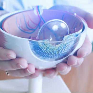
The EndoArt® implant (EyeYon Medical) is embedded through a clear corneal incision and positioned on the posterior stroma using an air-gas mixture.
“The implant looks like a contact lens—it is dome-shaped, 6.0 mm in diameter, and made of optically clear and flexible hydrophilic material that is bio-compatible and bio-stable. It works by preventing the transfer of aqueous humour into the cornea, thereby decreasing chronic corneal swelling,” Prof Auffarth said.
Importantly, the implant allows vital nutrients to reach the cornea on the periphery while blocking the harmful build-up of fluid in the centre.
Prof Auffarth presented some case studies of some of the first patients implanted with the device in Heidelberg, Germany, in June 2019.
“Both patients had very compromised corneas with severe pain deriving from bullous keratopathy after multiple surgeries, corneal decompensation, and failed DMEK. On postoperative day one, we could see clear corneas and a marked decrease in corneal thickness,” he said, adding the follow-up on both patients is now over 30 months and 36 months, respectively, and the corneas remain clear with no recurrence of oedema.
Looking at the analysis of the global data of EndoArt devices, Prof Auffarth said a total of 100 patients received implants to date with up to 36 months follow-up. For 22 patients, the implantation was performed as part of the first-in-human study, while for another 25 patients as part of the phase II study with a new surgical protocol.
Under the new protocol, 80% of 20 patients implanted needed just one rebubbling procedure or less, while 44% required zero rebubbling.
“We have learned a lot since the first cases were performed more than two years ago. The descemetorhexis needs meticulous endothelial removal with no tags or overlapping. We know now the optimal design of the implant is the 6.5 mm model in eyes with white-to-white corneal diameter greater than 10.8 mm.
Rebubbling is best performed with 10% PFP air-gas mixture. We also use a single 10/0 nylon suture and keep the patient supine for four hours postoperatively,” Prof Auffarth said, concluding the implant held a lot of promise considering the chronic shortage of donor tissue worldwide.
“With a device like this, there is no waiting list or eye bank needed for human tissue. It is safe, easy to implant, and can be removed or exchanged if necessary. The costs are lower than for conventional transplants, and it is a less traumatic surgery, which is obviously appealing to the patient,” he said. “It will be interesting to see what other expanded indications might enable this implant to be used in an ambulatory day care setting.”
Prof Auffarth gave this presentation at the 40th Congress of the ESCRS in Milan.
Gerd Auffarth MD, PhD, FEBO is Chairman of the Department of Ophthalmology at the Heidelberg University Eye Hospital and Head of the David J Apple Center for Vision Research, Heidelberg, Germany. Gerd.Auffarth@med.uni-heidelberg.de
Latest Articles
Nutrition and the Eye: A Recipe for Success
A look at the evidence for tasty ways of lowering risks and improving ocular health.
New Award to Encourage Research into Sustainable Practices
Sharing a Vision for the Future
ESCRS leaders update Trieste conference on ESCRS initiatives.
Extending Depth of Satisfaction
The ESCRS Eye Journal Club discuss a new study reviewing the causes and management of dissatisfaction after implantation of an EDOF IOL.
Conventional Versus Laser-Assisted Cataract Surgery
Evidence favours conventional technique in most cases.
AI Scribing and Telephone Management
Automating note-taking and call centres could boost practice efficiency.
AI Analysis and the Cornea
A combination of better imaging and AI deep learning could significantly improve corneal imaging and diagnosis.
Cooking a Feast for the Eyes
A cookbook to promote ocular health through thoughtful and traditional cuisine.
Need to Know: Spherical Aberration
Part three of this series examines spherical aberration and its influence on higher-order aberrations.
Generating AI’s Potential
How generative AI impacts medicine, society, and the environment.