Videos highlight ingenuity
Innovative techniques and new approaches showcased 2019 EURETINA Video Awards

Dermot McGrath
Published: Friday, September 6, 2019
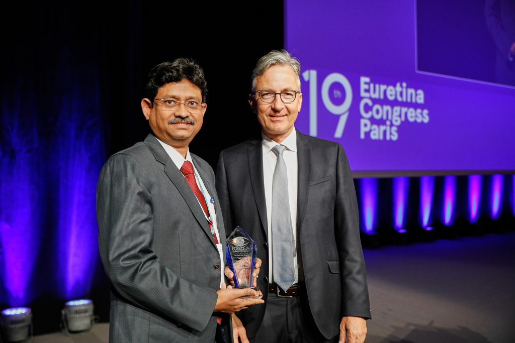 Rupak Kanti Biswas who won first prize in the EURETINA Video Competition Awards with Frank Holz, Incoming President, EURETINA[/caption]
AN innovative technique to close macular holes using a customised retinal graft and a new approach to treat the complete luxation of the crystalline lens while preserving the capsular bag were among some of the vitreoretinal surgical techniques showcased in the 2019 EURETINA Video Awards, with the very high standard of entries this year praised by Frank Holz, Incoming President of EURETINA.
First prize this year was awarded to Rupak Kanti Biswas MD of India for his entry on using an autologous retinal patch graft to close macular holes in cases where conventional methods such as ILM peeling or inverse flap technique have failed.
This year’s second prize was awarded to Fernando Gonzalez del Valle of Spain for his video “New vitreoretinal surgery to treat the complete luxation of the crystalline preserving the capsular bag”, while third prize went to Egor Arkhipov of Russia for “Multimodal approaches to the correction of post-traumatic eye changes”.
Dr Biswas’s prize-winning video entry presented an inventive approach to treating macular holes that had not been successfully closed using conventional surgical approaches. Rather than using autologous blood or lens capsular flap transplantation as a rescue strategy, Dr Biswas came up with the idea of plugging the macular hole with a customised patch of retinal tissue. Using a trephine that is 100 microns larger than the macular hole, Dr Biswas removes a patch of retinal tissue, which is then tucked into the hole using a soft-tipped cannula.
His hypothesis was that using a carefully sized patch of tissue would ensure end-to-end touch at the graft-host junction and facilitate neural regeneration. This was subsequently confirmed by multifocal ERG testing and automated perimetry assessment.
Dr Biswas has now performed seven cases using this technique with a minimal follow-up of one year and a maximum of 3.5 years.
“We have 70% of cases with the graft in place, the vision has improved in 40% of cases, and the rest of the patients had stable vision,” he said.
Dr Biswas noted that the key to success with this approach lies in harvesting the correct size of the retinal graft by trephination and not using any substance such as viscoelastic or perfluorocarbon which might hinder end to end touch.