The road to modernity
Transforming cataract extraction from couching to the world’s most frequent surgery

Howard Larkin
Published: Friday, December 6, 2019
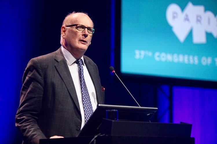 David Spalton delivering the 2019 ESCRS Heritage Lecture
Medical regulation stretches back in history preceding cataract surgery said Prof Spalton (Emeritus Professor of Ophthalmology, King’s College, London) in his introduction to the 2019 ESCRS Heritage Lecture “The Origins of Cataract Surgery”. The Code of Hammurabi, written in Mesopotamia, 1750 BC, lays down both a scale of fees and penalties too. “…if a physician open a tumour (over the eye) with an operating knife and saves the eye, he shall receive ten shekels…… if the physician makes a large incision with the operating knife and kills him or open a tumour with the operating knife and cut out the eye, his hands shall be cut off.” It has been suggested that the ‘tumour’ might mean a pterygium, if so there was a strong disincentive to be a corneal surgeon in the days of ancient Mesopotamia.
The development of cataract surgery reflects the advances in the understanding of the eye. References to ocular surgery date back to antiquity and couching cataracts – pushing the lens into the vitreous cavity to restore light perception – probably started in India some where about 1000–600 BC. The technology was probably introduced into Europe by the conquests of Alexander the Great (300 BC) and was the only treatment available until the introduction of extracapsular surgery by Daviel in 1745. Couching was performed in ancient Greece from about 300 BC, but early accounts of ocular surgery confuse corneal and lenticular pathologies and reflect no understanding of anatomy – Galen placed the lens anatomically in the centre of the eye and thought the lens was the organ of visual perception so that the eyes were thought to light up the world like car headlights, the extramission theory of vision. Prof Spalton explained “if you need the lens for visual perception, you were never going to take it out”. Instead, the ancient Greeks thought a cataract was a ‘hypochyma’, a dried up fluid lying in front of the lens and behind the iris.
The ideas of Galen and Celsius dominated thinking for 1,500 years. Celsius made the important observation that no perception of light was a bad prognostic indicator for surgical success. Another interesting observation was that Galen observed that “a goat suffering from hypochyma saw again when it fell and a thorn pierced its eye”, and it is interesting that a similar procedure – needling – was still the treatment of choice for congenital cataract when Prof Spalton was a resident at Moorfields in the 1970s, almost 2,000 years later.
By the 1500s there was still no understanding of the anatomy of the eye, its optics or the lens as the location of the cataract and it wasn’t until the taboo on human dissection started to be relaxed in the 16th Century that fundamental breakthroughs in understanding the anatomy and physiology of sight finally cleared the way for development of cataract surgery.
Understanding the lens
Vesalius in Padua published his major work on human anatomy (De Humani Corporis Fabrica; on the fabric of the human body) in 1543. He incorrectly still showed the lens as being at the centre of the eye but also showed that it was not the organ of perception. Bartisch in 1583 published the first textbook on ophthalmic surgery, at his own expense, with beautiful but gruesome detailed drawings of surgical procedures; he was trained by barber surgeons noting in the forward that “as he could not afford to go to school or university he had to restrict himself to surgery”! Forty years later Felix Platter’s anatomy placed the lens close to its true position and he identified the retina as the organ of visual perception. In 1619, Christophorus Scheiner accurately placed the lens and opened the sclera to observe the image on the retina.
Kepler was the father of modern optics, and in 1603 published his monumental book Astronomiae Pars Optica. He recognised the ocular image was projected inverted on the retina but the solution of this was of no interest to him as it was not optical – he was clearly a very focused individual – merely observing “that the image is corrected in the hollows of the brain by the activity of the soul”. Identification of the lens as the cataract surprisingly came rather later by Rolfinck in 1656 and confirmed in 1705 when Brisseau couched a soldier’s eye. He subsequently died and Brisseau extracted the lens, offering conclusive proof that the lens is the source of the cataract, Prof Spalton said.
Throughout this time couching was performed by itinerant surgeons such as the notorious ‘Chevalier’ John Taylor. He trained at St Thomas’ Hospital and travelled throughout Europe self styling himself as the oculist to the Pope and European royalty. Notably, he blinded Bach and possibly Handel too. It was said of him “bombast, effrontery, dishonesty, ignorance – all these qualities Taylor showed in perfection”.
Another interesting aside was that Rembrandt too painted a picture of Tobias and the angel operating on his father’s eye – careful study of which suggests that Rembrandt had watched couching surgery.
Birth of modern
cataract surgery
Against this background, the French surgeon Jacques Daviel (1693–1762) started to limit his practice to the eye in 1728, claiming to have performed more than 2,000 operations by 1737. In 1745 he opened the cornea of a patient with a curved needle and scissors to remove lens fragments and blood from a failed couching attempt, and the patient could immediately distinguish objects, though the eye was lost to infection.
Two years later, Daviel performed a couching procedure on M Garion, a wig maker. He was unable to depress the lens and decided to operate as he had done on the previous patient, publishing an account in 1753. By 1756 Daviel reported 434 extractions with 88% “success”, a marked improvement over couching. All procedures were done of course without anaesthetic, leading Daviel to observe: “Stabilisation is a problem… There is general unsteadiness of the globe from the influence of the mind on the organ about to be injured.” A remarkable turn of phrase, Prof Spalton commented.
The popularity of lens extraction and its requirement for quick and accurate surgery led to a blossoming proliferation of keratomes, specula and other instruments, while controversy swirled over whether couching or lens extraction was best. Refinements from many surgeons accelerated through the 19th Century, Albrecht von Graefe being a notable contributor, and there are many instruments that are similar to what we use today. The invention of cocaine for topical anaesthesia by Dr Karl Koller in Vienna in 1884 was a major contribution that did much to rapidly advance surgery.
While the extracapsular technique pioneered by Daviel and continuously improved was safe and relatively easy, it was limited to mature cataracts and subject to secondary membrane development and inflammation. Through the first half of the 20th Century it gave way to intracapsular techniques that, while more technically challenging and requiring larger incisions, were suitable for less mature cataracts. Pioneering advances included manual expression of the lens by Col Henry Smith in India, alpha chymotrypsin to dissolve zonules by Jose Barraquer and cryoextraction by Krawicz and Kelman. However, it was the invention of the intraocular lens by Sir Harold Ridley followed by phacoemulsification by Charles Kelman that made cataract surgery the most widely performed and highly successful procedure it is today.
Many surgeons made great contributions, but Daviel was the first of the four great pioneering surgeons followed by von Graefe, Ridley and Kelman, David Spalton concluded, but we have run out of time and the story
of their contributions will have to be
the subject of a Heritage Lecture
another day.
The complete lecture is available on ESCRS on Demand on the ESCRS website.
David Spalton: profspalton@gmail.com
David Spalton delivering the 2019 ESCRS Heritage Lecture
Medical regulation stretches back in history preceding cataract surgery said Prof Spalton (Emeritus Professor of Ophthalmology, King’s College, London) in his introduction to the 2019 ESCRS Heritage Lecture “The Origins of Cataract Surgery”. The Code of Hammurabi, written in Mesopotamia, 1750 BC, lays down both a scale of fees and penalties too. “…if a physician open a tumour (over the eye) with an operating knife and saves the eye, he shall receive ten shekels…… if the physician makes a large incision with the operating knife and kills him or open a tumour with the operating knife and cut out the eye, his hands shall be cut off.” It has been suggested that the ‘tumour’ might mean a pterygium, if so there was a strong disincentive to be a corneal surgeon in the days of ancient Mesopotamia.
The development of cataract surgery reflects the advances in the understanding of the eye. References to ocular surgery date back to antiquity and couching cataracts – pushing the lens into the vitreous cavity to restore light perception – probably started in India some where about 1000–600 BC. The technology was probably introduced into Europe by the conquests of Alexander the Great (300 BC) and was the only treatment available until the introduction of extracapsular surgery by Daviel in 1745. Couching was performed in ancient Greece from about 300 BC, but early accounts of ocular surgery confuse corneal and lenticular pathologies and reflect no understanding of anatomy – Galen placed the lens anatomically in the centre of the eye and thought the lens was the organ of visual perception so that the eyes were thought to light up the world like car headlights, the extramission theory of vision. Prof Spalton explained “if you need the lens for visual perception, you were never going to take it out”. Instead, the ancient Greeks thought a cataract was a ‘hypochyma’, a dried up fluid lying in front of the lens and behind the iris.
The ideas of Galen and Celsius dominated thinking for 1,500 years. Celsius made the important observation that no perception of light was a bad prognostic indicator for surgical success. Another interesting observation was that Galen observed that “a goat suffering from hypochyma saw again when it fell and a thorn pierced its eye”, and it is interesting that a similar procedure – needling – was still the treatment of choice for congenital cataract when Prof Spalton was a resident at Moorfields in the 1970s, almost 2,000 years later.
By the 1500s there was still no understanding of the anatomy of the eye, its optics or the lens as the location of the cataract and it wasn’t until the taboo on human dissection started to be relaxed in the 16th Century that fundamental breakthroughs in understanding the anatomy and physiology of sight finally cleared the way for development of cataract surgery.
Understanding the lens
Vesalius in Padua published his major work on human anatomy (De Humani Corporis Fabrica; on the fabric of the human body) in 1543. He incorrectly still showed the lens as being at the centre of the eye but also showed that it was not the organ of perception. Bartisch in 1583 published the first textbook on ophthalmic surgery, at his own expense, with beautiful but gruesome detailed drawings of surgical procedures; he was trained by barber surgeons noting in the forward that “as he could not afford to go to school or university he had to restrict himself to surgery”! Forty years later Felix Platter’s anatomy placed the lens close to its true position and he identified the retina as the organ of visual perception. In 1619, Christophorus Scheiner accurately placed the lens and opened the sclera to observe the image on the retina.
Kepler was the father of modern optics, and in 1603 published his monumental book Astronomiae Pars Optica. He recognised the ocular image was projected inverted on the retina but the solution of this was of no interest to him as it was not optical – he was clearly a very focused individual – merely observing “that the image is corrected in the hollows of the brain by the activity of the soul”. Identification of the lens as the cataract surprisingly came rather later by Rolfinck in 1656 and confirmed in 1705 when Brisseau couched a soldier’s eye. He subsequently died and Brisseau extracted the lens, offering conclusive proof that the lens is the source of the cataract, Prof Spalton said.
Throughout this time couching was performed by itinerant surgeons such as the notorious ‘Chevalier’ John Taylor. He trained at St Thomas’ Hospital and travelled throughout Europe self styling himself as the oculist to the Pope and European royalty. Notably, he blinded Bach and possibly Handel too. It was said of him “bombast, effrontery, dishonesty, ignorance – all these qualities Taylor showed in perfection”.
Another interesting aside was that Rembrandt too painted a picture of Tobias and the angel operating on his father’s eye – careful study of which suggests that Rembrandt had watched couching surgery.
Birth of modern
cataract surgery
Against this background, the French surgeon Jacques Daviel (1693–1762) started to limit his practice to the eye in 1728, claiming to have performed more than 2,000 operations by 1737. In 1745 he opened the cornea of a patient with a curved needle and scissors to remove lens fragments and blood from a failed couching attempt, and the patient could immediately distinguish objects, though the eye was lost to infection.
Two years later, Daviel performed a couching procedure on M Garion, a wig maker. He was unable to depress the lens and decided to operate as he had done on the previous patient, publishing an account in 1753. By 1756 Daviel reported 434 extractions with 88% “success”, a marked improvement over couching. All procedures were done of course without anaesthetic, leading Daviel to observe: “Stabilisation is a problem… There is general unsteadiness of the globe from the influence of the mind on the organ about to be injured.” A remarkable turn of phrase, Prof Spalton commented.
The popularity of lens extraction and its requirement for quick and accurate surgery led to a blossoming proliferation of keratomes, specula and other instruments, while controversy swirled over whether couching or lens extraction was best. Refinements from many surgeons accelerated through the 19th Century, Albrecht von Graefe being a notable contributor, and there are many instruments that are similar to what we use today. The invention of cocaine for topical anaesthesia by Dr Karl Koller in Vienna in 1884 was a major contribution that did much to rapidly advance surgery.
While the extracapsular technique pioneered by Daviel and continuously improved was safe and relatively easy, it was limited to mature cataracts and subject to secondary membrane development and inflammation. Through the first half of the 20th Century it gave way to intracapsular techniques that, while more technically challenging and requiring larger incisions, were suitable for less mature cataracts. Pioneering advances included manual expression of the lens by Col Henry Smith in India, alpha chymotrypsin to dissolve zonules by Jose Barraquer and cryoextraction by Krawicz and Kelman. However, it was the invention of the intraocular lens by Sir Harold Ridley followed by phacoemulsification by Charles Kelman that made cataract surgery the most widely performed and highly successful procedure it is today.
Many surgeons made great contributions, but Daviel was the first of the four great pioneering surgeons followed by von Graefe, Ridley and Kelman, David Spalton concluded, but we have run out of time and the story
of their contributions will have to be
the subject of a Heritage Lecture
another day.
The complete lecture is available on ESCRS on Demand on the ESCRS website.
David Spalton: profspalton@gmail.com