Cornea
PDEK: A Unique Mix of Anatomy and Technique
The first in a new series on pre-Descemet endothelial keratoplasty examines the anatomical and surgical factors behind its low rebubbling rates.


Soosan Jacob
Published: Thursday, November 5, 2026
“ PDEK offers the ease of handling associated with DSAEK while retaining many optical advantages of DMEK. “
Endothelial keratoplasty (EK) has evolved into three major surgical options: Descemet stripping automated endothelial keratoplasty (DSAEK), Descemet membrane endothelial keratoplasty (DMEK), and pre-Descemet endothelial keratoplasty (PDEK). Each procedure replaces dysfunctional corneal endothelium, but the transplanted tissue layers, surgical handling, and postoperative graft behaviour differ significantly.
To understand these differences, it is necessary to revisit corneal anatomy. In 2013, Professor Harminder Dua described an additional layer between the posterior stroma and Descemet’s membrane (DM), now called the pre-Descemet’s layer (PDL), or Dua’s layer. This layer is central to understanding PDEK surgery.
Comparing different EK procedures
In DSAEK, transplanted tissue includes endothelium, DM, PDL, and a variable thickness of posterior stroma. The stromal component varies from approximately 150 microns to 80 or even 50 microns, depending on whether standard DSAEK, ultrathin DSAEK, or nano-DSAEK is being performed. Though posterior stroma makes the graft relatively easier to handle, it also results in transplantation of a thicker graft. It therefore increases effective postoperative corneal thickness, requires the endothelium to pump fluid across a thicker corneal complex, and affects refraction, visual quality, and rejection rates.
DMEK was therefore a major refinement because only the DM and endothelium were transplanted. However, it brought its own challenges. At only approximately 15 microns thick, DMEK produces excellent optical results. Its extremely thin, delicate nature and tendency to scroll tightly, however, make surgery difficult, especially in challenging anterior segments. DMEK graft preparation is also more difficult with young donors, particularly those younger than 40 to 50 years of age, as the graft scrolls even tighter. As a result, DMEK grafts are often preferred from older donors, although they generally have lower endothelial cell counts than younger ones. Additionally, in diabetic donors, the risk of tissue preparation failure is higher. Thus, while DMEK is anatomically superior, it may not always allow the use of the youngest and potentially healthiest endothelial tissue.
PDEK occupies a middle ground. The PDEK graft includes the PDL, DM, and endothelium. It is slightly thicker than DMEK, since it is approximately 25 microns rather than 15 microns, but still far thinner than DSAEK. Its major advantage is that it can be prepared from donors of any age. In practice, PDEK grafts are often taken from donors younger than 40 years old and frequently from those younger than 20 or even 10 years, because younger tissue generally has healthier endothelium and higher endothelial cell counts.
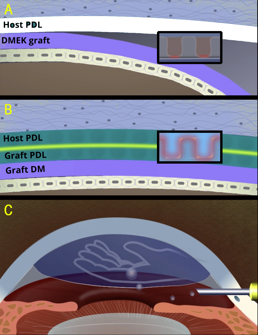
The PDL gives PDEK distinctive surgical characteristics. It is an acellular, tough layer, about 10 to 15 microns thick, made of compact type I collagen lamellae arranged in longitudinal, transverse, and oblique directions. While its elastin content gives the graft elasticity, the PDL gives it a splinting effect, and the collagen lamellae make it resistant to tearing. Therefore, although PDEK grafts may still scroll like DMEK grafts, the PDL supports the DM and endothelium, making the graft easier to manipulate within the anterior chamber (AC). In this sense, PDEK offers the ease of handling associated with DSAEK while retaining many optical advantages of DMEK.
Rebubbling
A crucial advantage of PDEK is its low detachment rate of about 3%. DMEK has a reported graft detachment rate ranging from 2% to 82%, with a rough average of 33%. Thus, the need for postoperative rebubbling is significantly lower with PDEK.
Rebubbling, whether performed in the operating room or clinic, exposes the patient to a second intervention and additional risks, including infection, excess air fill, pupillary block, Urrets-Zavalia syndrome, anterior capsular cataract, repeat detachment, underfill, and hypotony. Higher rebubbling rates also add significant burden in terms of cost, delayed visual recovery, and lost time from work. PDEK, in the author’s own experience, has a lower detachment rate even in difficult eyes and complex anterior chambers, which removes postoperative uncertainty for both patient and surgeon.
Why should PDEK detach less often?
The answer to this key question has a few proposed hypotheses.
One proposed explanation again returns to the PDL. DM is composed mainly of type IV and type VIII collagen. Type IV collagen forms a loose, two-dimensional meshwork or ‘chicken-wire’ network, specialised for basement membrane function, endothelial support, selective permeability, cellular attachment, and migration. Type VIII collagen forms a short, non-fibrillar hexagonal network that contributes to DM structure. In contrast, type I collagen, which forms the PDL, is a fibrillar collagen that provides tensile strength and structural toughness.
In DMEK, the donor DM and endothelium are placed against the recipient’s posterior corneal surface after removal of host DM and endothelium. Thus, the donor type IV and type VIII collagen complex must adhere to the host posterior stromal or PDL surface. This may create a potential plane or space at the interface, contributing to detachment.
In PDEK, especially when prepared from young donors, the PDL, DM, and endothelium remain tightly attached as one complex. In younger donors, the bond between PDL and DM is stronger, making spontaneous separation less likely. During surgery, the host DM and endothelium are removed, and the donor PDL–DM–endothelium complex is placed against the host. This results in donor PDL apposing the host PDL. Since both are similar tissues and have relatively rough surfaces, this ‘rough to rough’ apposition may provide more secure adherence than the smoother DM to PDL interface in DMEK. It may explain the lower rebubbling rate observed with PDEK.
The air pump-assisted PDEK technique, as previously described, may further reduce detachment rates. During surgery, pressurised air infusion maintains the AC and acts like a third hand. It helps displace fluid trapped between the graft and the host surface, even without venting incisions. Second, it allows the surgeon to identify and remove even small edge folds, which are common starting points for graft detachment. Third, it improves graft centration, placing the graft in an ideal anatomical location that promotes attachment. Fourth, the small period of higher intraoperative pressure eliminates the need to close the eye with a full air bubble followed by later burping to avoid pupillary block. With air pump-assisted PDEK, the graft is already well attached by the time the eye is closed, and an approximate 80% air bubble is generally sufficient. This has also completely negated the need for long-acting gas.
The air pump-assisted technique is also particularly helpful in complex anterior chambers. For instance, in eyes with intracameral bleeding, the pressurised air can tamponade bleeding, float the graft quickly, and still allow blood washout, even after graft floatation. This reduces postoperative fibrin, which could otherwise contract and contribute to graft detachment. Similarly, the air pump can support the graft while additional manoeuvres are performed, such as synechiolysis, pupilloplasty, vitrectomy, or other intraocular steps. It also allows rapid unfolding in complex eyes even with peripheral edge folds present, which can then be unfolded using the air pump.
Thus, the low rebubbling rate in PDEK is explained by a combination of graft anatomy, donor selection, interface behaviour, and surgical technique. The PDEK graft is thin enough to offer DMEK-like optical advantages, strong enough to provide DSAEK-like handling, biologically suited to using younger donor tissue and has anatomy that promotes attachment. When combined with the air pump-assisted technique, it provides stable intraoperative apposition and lower postoperative detachment.
Dr Soosan Jacob is Director and Chief of Dr Agarwal’s Refractive and Cornea Foundation at Dr Agarwal’s Eye Hospital, Chennai, India, and can be reached at dr_soosanj@hotmail.com.