“The eye is an approachable part of the brain.” This quotation is the premise on which half of the subject matter of this handbook is based. The other premise, that optical coherence tomography (OCT) can be used to help diagnose causes of reduced visual acuity not only in adults, but also in children and even infants, forms an intriguing introduction to this 330-page handbook.
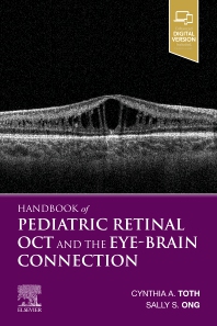
The Handbook of Pediatric Retinal OCT & the Eye-Brain Connection (Elsevier), edited by Cynthia Toth and Sally Ong, might be the first of its kind. Professor Toth has been imaging infants for more than 15 years, and has published extensively on the subject, sharing her insights on OCT for retinopathy of prematurity, nystagmus and ocular albinism, among others.
The first part of the handbook introduces paediatric retinal OCT imaging, informing the reader on how to organise the logistics of such imaging and how to analyse the resulting scans. Because paediatric retinas are not simply smaller versions of the adult structure, section 2 delves into “Age-dependent Features & Common Abnormalities”. Examples include a fascinating discussion of foveal development from 30 weeks of gestation to childhood and a surprisingly large range of vitreoretinal abnormalities.
After having covered the practical matters of OCT imaging and interpretation, the authors turn to the specific diseases that will be encountered in clinical practice. What does Best disease look like on OCT in a six-year-old girl? How about FEVR in a five-year-old? It’s all here. Each disorder is illustrated by the highest-quality OCT scans and ancillary imaging, mostly courtesy of the Duke Imaging Center.
What about the eye-brain connection? “Pediatric intermediate uveitis may be rarely associated with multiple sclerosis,” and “Torpedo maculopathy has been reported in one patient with tuberous sclerosis” are some examples.
This handbook is intended for retinal specialists, ambitious paediatric ophthalmologists and ophthalmology departments in hospitals with referral centres for neonatal intensive care and/or complex paediatric disease. With its highly organised structure and clear overview of rarer retinal disease, it can also help the ready study for large examinations.