Refractive Surgery
Patient Portal
Refractive surgery is functional, not cosmetic, and can reduce dependence on spectacles and contact lenses. However, it does share many similarities with cosmetic surgery; for most patients, refractive surgery is elective and self-funded and is predominantly provided by the private healthcare sector.
Your questions about Refractive Surgery
Checklist for Patients
For use in consultation with your surgeon.
Think carefully before having refractive surgery. Take this checklist to your consultation with your refractive surgeon performing the procedure. Discuss each item with your surgeon to help you make the decision that is right for you before having refractive surgery.
Meet the eye surgeon who will be performing your procedure.
Choose your surgeon and clinic carefully.
Get information on all the planned and possible costs. The cost of your surgery will depend on the type of procedure you have agreed with your surgeon. You should find out ALL the costs involved and what your rights are to refunds/return of deposits if you change your mind after you have paid some or all of the costs. Although a fee may be charged for your initial consultation, check if the deposit for surgery is fully refundable within a reasonable time period if you choose not to proceed. Your procedure fee will normally include follow up clinic visits and treatment for any problems resulting from surgery. Ask how long after the surgery you are covered for free additional treatment.
Tell the eye surgeon performing the procedure what you want to change about your vision and why. Let them know what you would consider to be a successful outcome.
Discuss the possible benefits and risks of all treatment options and find out if the surgeon can give you the results you want. After reviewing your test results and examining your eyes, the operating surgeon should advise you in person about the best procedure choice for you and take your consent.
Find out what the procedure will involve, the likely results, when you will see them and how long they will last. Will surgery need to be repeated in the future? Where and when will the procedure take place and how long will it take? Ask your surgeon what kind of activities might still require use of glasses after surgery.
Like any surgical procedure there are risks as well as benefits to consider. Find out what could go wrong with the procedure you want, how likely is it to happen and what can be done to correct it.
Ask your surgeon what the most common complications have been in their experience for this procedure, how often these have occurred and what the surgeon did to correct them. Consider how you would cope if anything went wrong.
Find out what to expect after the surgery including recovery time, how long any pain is likely to last and what you will and will not be able to do after surgery.
Find out what follow-up care will be needed after the surgery. Who will do it and where will it be done. Ask what your aftercare package will and will not cover, how you will be looked after and by whom, and how long the surgeon and hospital or clinic will continue to support you. What will happen if something does not go to plan and who will pay? What will happen if something goes wrong immediately afterwards or if you are not happy with your outcome? Is there immediate access to care? More commonly, problems can be corrected with changes in medication or additional surgery. Typically, these additional operations feel like the original surgery and have a similar recovery period.
Loss of vision
Permanent serious loss of vision is rare after laser vision correction. In the worst likely scenario, a form of corneal transplantation may be required to replace a damaged block of tissue in the cornea. Problems that can lead to the need for transplantation include scarring after infection or an abnormal healing response, and an unstable corneal shape – also called corneal ectasia. These problems occur infrequently and can often be corrected without transplant surgery. Less than 1 in 5000 patients require a corneal transplant to restore vision after laser surgery, and good vision can normally be restored when transplantation is necessary although glasses or contact lenses may be required.
Additional surgery
Much more commonly, a second operation is needed to correct a problem occurring at the time of surgery or afterwards. This would normally be a procedure that feels similar to the original laser vision correction, with a similar recovery period. Some of these procedures need to be repeated. If you have a problem, your surgeon will explain what it is and why further surgery is required. Up to 1 in 10 patients require some form of additional surgery in order to get the best result.
Before you have surgery, make sure you have the contact details of a named doctor who can deal with any problems after the procedure.
- Don’t be afraid to ask questions
- Never feel rushed or pressured into giving your consent to have surgery by special offers that are for a limited time only or any discounts in price eg two-for-one deals
- Only the operating surgeon should advise you about the procedure choice and take your consent for surgery
- You can change your mind at any point and you can ask for a second opinion from another surgeon
Take at least a week after your consultation with the surgeon performing the procedure to think things through before surgery.
Accommodation the reflex by which the eye brings a near object into focus by changing the shape of the natural lens. The natural lens gets less flexible with age, and powers of accommodation diminish. This affects reading vision and the ability to compensate for hyperopia.
Artisan/ArtiflexPIOL this is a type of PIOL that is clipped on to the iris during implantation. PIOLs are a commonly used alternative to laser vision correction for younger patients.
Astigmatism irregular defocus, or an eye that is ‘more rugby ball shaped than football shaped.’ The first number in your prescription for glasses describes the amount of long or short sight. The second number describes the amount of astigmatism. Most of us have at least some astigmatism, and a small amount can even help extend the range of activities you can do without reading glasses when you get older.
Biometry this test involves measurement of the eye and a set of calculations (biometry formulae) that help determine the right focusing power for an IOL before cataract surgery or RLE.
Binocular vision this is vision measured with the two eyes open.
Cataract when the natural lens gets misty enough to make vision hazy, it is called a cataract.
Cataract surgery this is surgery to replace the natural lens with an IOL. Cataract surgery is identical to RLE but is performed in patients who cannot see clearly despite using glasses or contact lenses with the main aim of restoring clear vision.
Conjunctiva the membrane covering the white of the eye. The conjunctiva produces mucus to help spread the watery tear film over the eye surface.
Cornea the clear part of the eye wall at the front of the eye. Two thirds of the focusing power of the eye is in the cornea.
Corneal epithelium the corneal skin layer
Corneal topography this is a scan that maps the surface curvature of the cornea.
D or Dioptre a unit for measuring the refractive power of a lens.
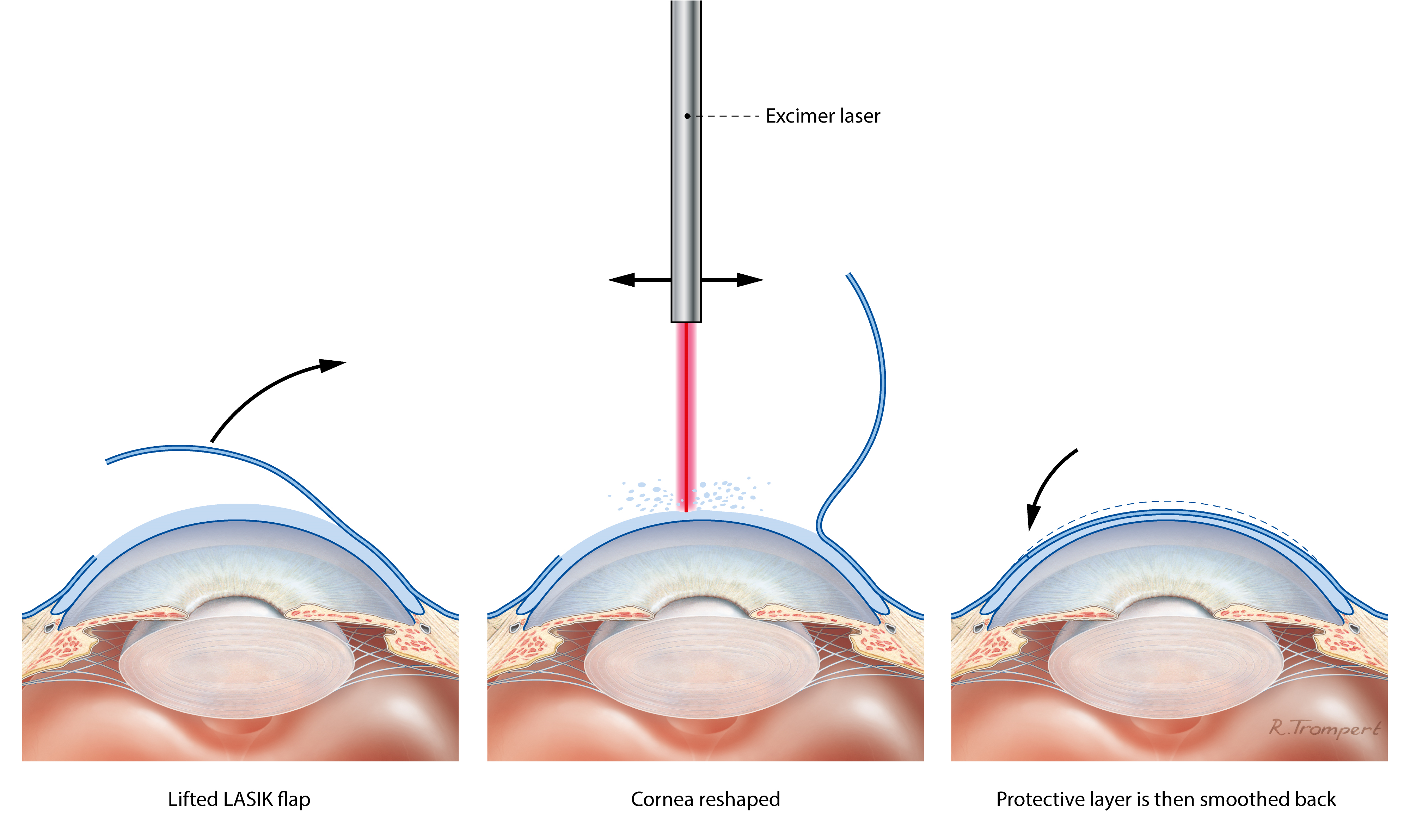
Excimer laser this type of laser removes tissue by non-thermal vaporisation (photoablation). Excimer lasers are extremely accurate and do not damage the surrounding tissues. They are used in LASIK and surface laser treatments (PRK, LASEK and TransPRK).
Femtosecond laser this type of laser is designed to cut any 3D shape in clear eye tissues such as the natural lens or the cornea with a high degree of accuracy. They work by creating a 3D pattern of tiny gas bubbles, which is traced through the target tissue at high speed. Femtosecond lasers are used in LASIK, SMILE, and increasingly in RLE and Cataract surgery.
Floaters floating shadows cast on the retina by opacities in the vitreous. Most of us are aware of floaters in some lighting conditions.
Glaucoma this is a condition in which the optic nerve is gradually damaged causing the visual field to contract. Left untreated, patients with glaucoma may develop ‘tunnel vision.’ Glaucoma is often associated with a higher than normal intraocular pressure, and treatment is centred on drugs or surgery to lower the intraocular pressure.
Hyperopia long sight. People with hyperopia typically have good vision as young adults. As they get older, they find themselves reliant on glasses for
reading, and then for the distance vision too. Younger people are able to compensate for hyperopia by accommodation. The amount of hyperopia is shown by a positive number in your spectacle prescription.
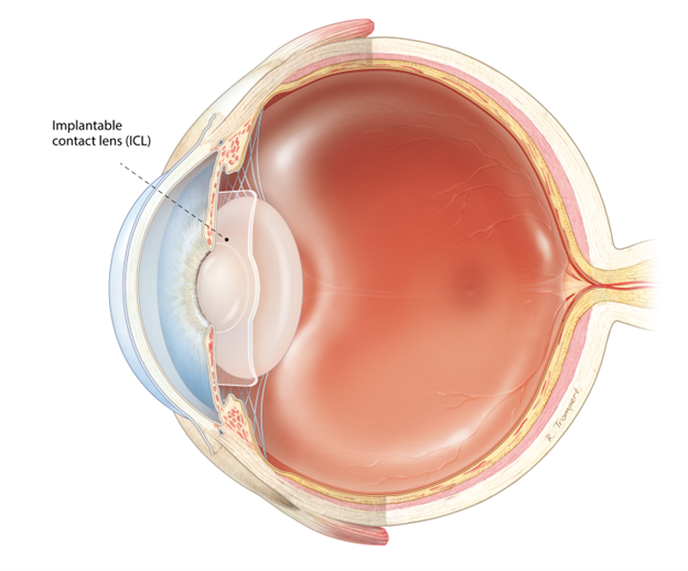
ICL (implantable contact lens) this is the most commonly used type of PIOL. It is implanted behind the iris and vaults over the natural lens – a bit like a contact lens implanted in the eye. ICL implantation is a commonly used alternative to laser vision correction in younger patients.
IOL (intraocular lens) IOLs are small synthetic lens implants that are used to replace the natural lens in cataract surgery and RLE.
Intraocular pressure (IOP) this is the pressure of fluid within the eye. It is often measured with a puff of air in routine eye checks or (more accurately) with a yellow drop and a blue light.
Iris this is the coloured part of the eye behind the cornea that expands and contracts in response to light to dilate or constrict the pupil.
LASEK (laser assisted sub-epithelial keratectomy) this is a form of surface laser treatment in which the corneal skin layer is soaked with dilute alcohol to loosen it before removal.
Laser vision correction correction of sight using excimer and/or femtosecond lasers to alter the curvature and focusing power of the cornea.
LASIK (laser in situ keratomileusis) this is the commonest form of laser vision correction in which a thin protective flap is created using a femtosecond laser. The protective flap is moved aside by the surgeon before optical reshaping of the cornea using an excimer laser. The flap is then replaced, and adheres without stitches, keeping the corneal skin layer intact and giving a fast visual recovery.
Meibomian glands the specialized oil glands in the eyelids that pump out a stabilizing layer of oil each time we blink that floats on top of the watery layer of the tear film. Inflammation of the eyelid margins upsetting this layer (blepharitis or meibomitis) is one of the commonest reasons for eye discomfort.
Micromonovision this is the name often given to the strategy of aiming for a clearer distance focus in one eye and a clearer focus at arms’ length in the other. Input from the two eyes combines to extend the range of focus for patients in the reading glasses age group undergoing vision correction surgery.
Monofocal IOL an IOL with one clear point of focus. These are the lenses most commonly used in standard cataract surgery. They have fewer optical side effects than multifocal lenses, but glasses are normally required for at least some activities after implantation.
Multifocal IOL an IOL with more than one point of clear focus This includes enhanced monofocal and Extended Depth of Focus lenses. Multifocal IOLs are often used in RLE in order to help increase freedom from glasses in the near range as well as providing good distance vision.
Myopia short sight. People with myopia are able to see up close but not in the distance. They typically first need glasses as school age children. The amount of myopia is shown as a negative number in your spectacle prescription.
Natural Lens the natural lens sits just behind the pupil and is suspended by a trampoline like array of microligaments from the ciliary muscle, which contracts during accommodation. The natural lens accounts for one third of the focusing power of the eye and is the flexible element of focus. The natural lens gets less flexible with age. It also becomes less clear as we get older. If the natural lens gets misty enough to make vision hazy, it is called a cataract.
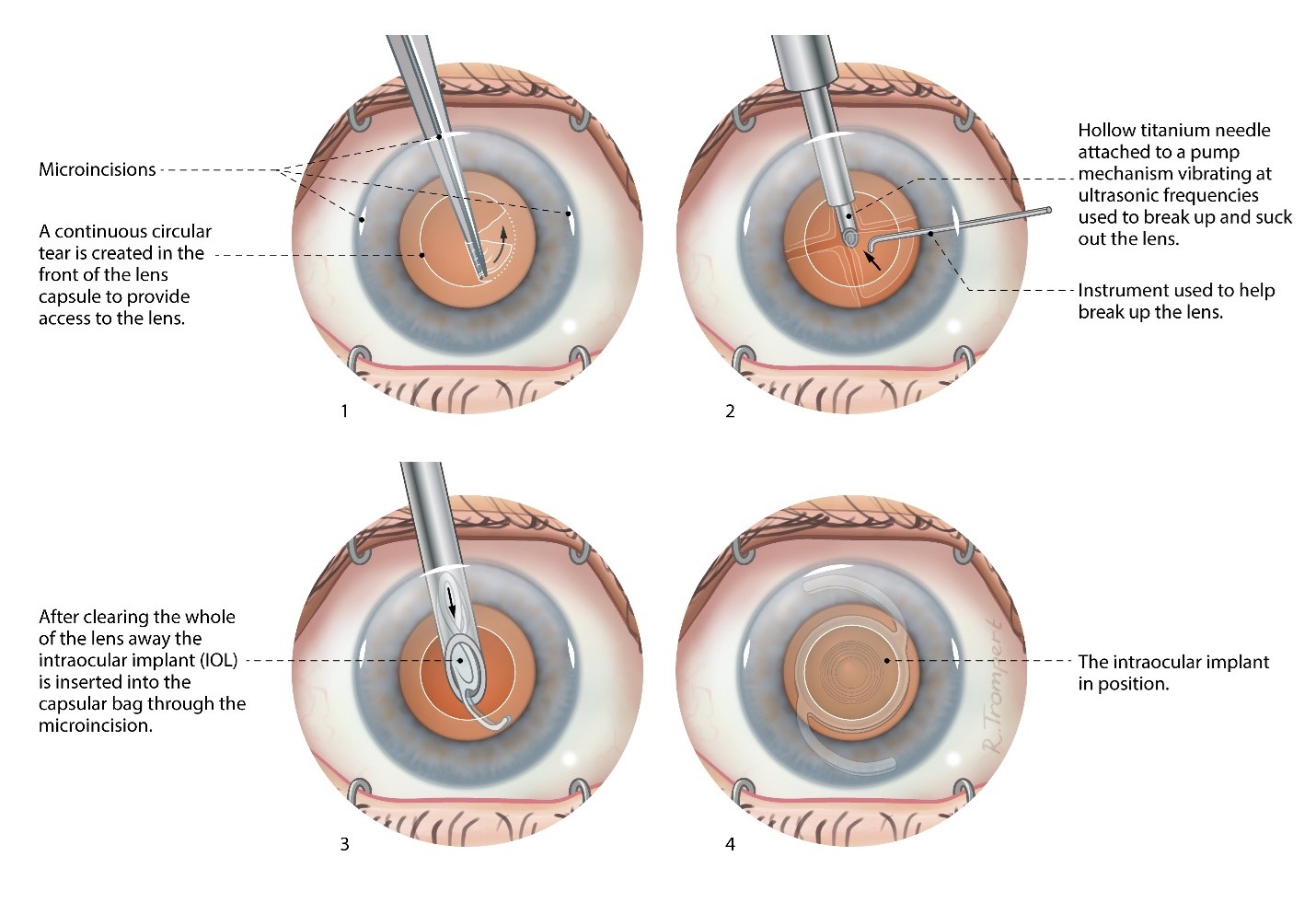
Phacoemulsification this is the standard technique for liquefying the natural lens in cataract surgery and RLE. Energy delivered at ultrasonic frequencies from the tip of a fine, hollow probe liquefies the lens. Fluid is washed continuously into the eye around the probe, and the liquefied lens material is sucked away. Where previously the natural lens had to be shelled out like a pea, phacoemulsification allows it to be removed through a tiny, key-hole entry into the eye. This development revolutionised modern cataract surgery. Femtosecond lasers are now commonly used to break the lens up into small fragments before phacoemulsification.
Posterior capsule opacification (PCO) in cataract surgery and RLE, the IOL is implanted within the capsule of the natural lens. This thin, clear membrane then shrink-wraps the IOL and stabilises it in the natural position in the eye. As part of this healing up process, the membrane often goes misty, causing gradual loss of vision after surgery. This is posterior capsule opacification (PCO). It can be treated successfully with a one-off minor laser procedure called YAG capsulotomy.
Presbyopia age related loss of reading vision and the ability to focus on a near object without help from glasses.
PIOL (phakic intraocular lens) an eye that still has the natural lens in place is described as ‘phakic.’ PIOLs are small, synthetic lenses that are implanted in the eye without taking the natural lens out. PIOL implantation is an alternative to laser vision correction for younger patients.
Posterior vitreous detachment (PVD) as we age, the vitreous gel shrinks and will often peel off the back of the eye. For some of us, this event passes unnoticed. In others, a new shower of floaters will prompt them to attend for an eye examination. Doctors examining patients after a PVD look carefully for any abnormal attachment of the gel to the retina which could lead to a retinal detachment.
PRK (photorefractive keratectomy) this is a form of surface laser treatment in which preparatory removal of the corneal skin layer is done by the surgeon.
Refraction this is the test that is done to determine the numbers in your spectacle prescription and the amount of myopia, hyperopia or astigmatism.
Refractive surgery this is another name for vision correction surgery or surgery to reduce the need for glasses and contact lenses.
Retina the carpet of light sensitive cells lining the back of the eye, which is sometimes described as ‘the film in the camera.’ Images are focused on the retina by the cornea, the natural lens. Information from the retina is fed through the optic nerve to the visual areas of the brain.
Retinal detachment the retina sometimes detaches from the eye wall and its blood supply. Urgent surgery is then required to re-attach the retina in order to prevent visual loss. Patients with high myopia are more likely to get a retinal detachment. Retinal detachments are also more common in the early years after cataract surgery or RLE. Warning signs are a sudden change in vision with field loss (a dark shadow in part of the visual field); flashing lights (arcs of light – even with the eyes closed); and a sudden new shower of floaters.
RLE (refractive lens exchange) this is surgery to replace the natural lens with an IOL. RLE is identical to cataract surgery but is performed in patients who can see clearly if they wear glasses or contact lenses. The aim of RLE is to help people to see clearly for more activities without glasses or contact lenses.
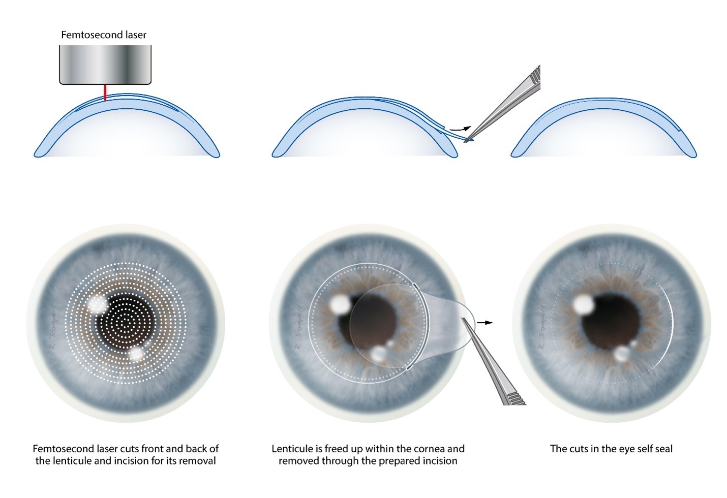
SMILE (small incision lenticule extraction) this is a form of laser vision correction in which a lens shaped piece of corneal tissue is marked out using a femtosecond laser and removed surgically through a small incision.
Surface laser treatment a collective term for PRK, LASEK, TransPRK and other similar forms of laser vision correction in which optical reshaping of the cornea is performed on the corneal surface after removal of the corneal skin layer.
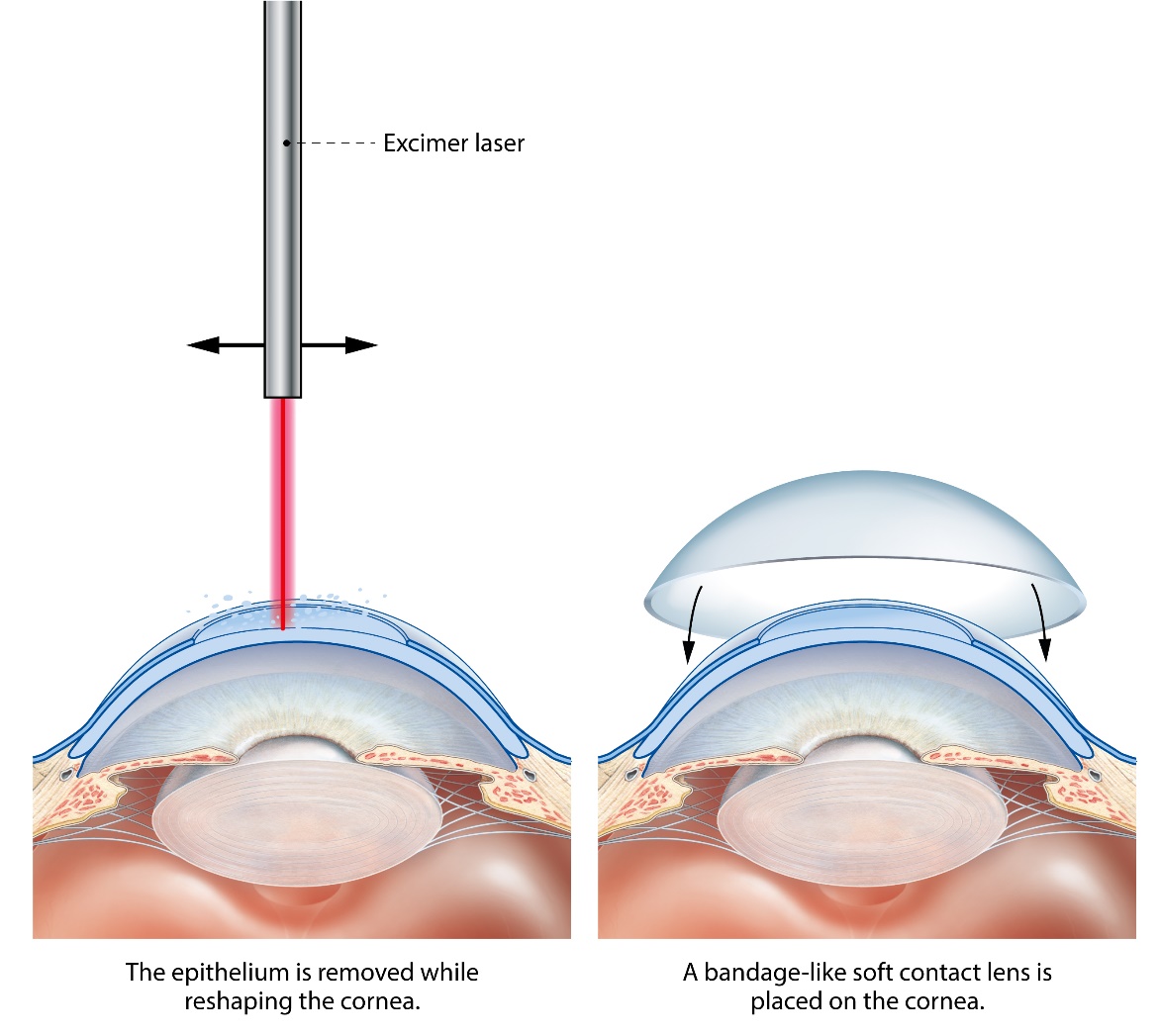
TransPRK (transepithelial PRK) this is a form of surface laser treatment in which the corneal skin layer is removed by the laser itself.
Tear film a multilayered wet film covering the front of the eye, which is essential for vision and comfort. A layer of mucus produced by specialized cells in the conjunctiva helps the watery layer of the tear film to spread over the eye. The watery layer is stabilized between blinks by an oily layer that forms a thin film (a bit like petrol floating on water) and acts to prevent localized evaporation. The oily layer is pumped out from specialized glands in the eyelids called meibomian glands each time we blink.
Visual acuity this is the main measurement of how well we can see and is tested by asking you to read down a chart which has smaller letters on each line. The lower you can read, the better your visual acuity is. Measurements are expressed as a fraction. If your visual acuity is 6/6 (normal) then you can see at 6 metres what a normally sighted person can see at 6 metres. If your vision is 6/9 you can see at 6 metres what a normally sighted person can see from 9 metres away et cetera. In the USA, the same measurement is made in feet, with 6 metres being roughly 20 feet. Many people are familiar with the American definition of normal ‘20/20’ vision. This is the same as 6/6 vision in the UK measurement.
Vitreous this is the gel filling the back of the eye. It tends to shrink as we age and accumulates wrinkles and opacities that cast floating shadows (floaters) on the retina.
Wavefront scan this is an optical map of your eye used to guide modern excimer laser treatments.
YAG capsulotomy a one-off minor laser procedure used to treat posterior capsule opacification (PCO).
Laser Vision Correction
Modern surgical lasers are able to alter the curvature and focusing power of the front surface of the eye (the cornea) very accurately to correct short sight (myopia), long sight (hyperopia), and astigmatism.
Three types of procedure are commonly used: LASIK, surface laser treatments (PRK, LASEK, TransPRK) and SMILE. Risks and benefits are similar, and all these procedures normally produce very good results in the right patients. Differences between these laser vision correction procedures are explained below. If you are suitable for laser vision correction, your surgeon will discuss which type of procedure is the best option for you.
For most patients, vision after laser correction is similar to vision in contact lenses before surgery, without the potential discomfort and limitations on activity. Glasses may still be required for some activities after treatment, particularly for reading in older patients. Over 95% of patients are satisfied with the outcome of surgery, and many describe it as life changing.
Although laser vision correction is often bracketed with cosmetic surgery procedures, the benefits are mainly functional. It is designed to make you less dependent on glasses and contact lenses, helping you to lead an active lifestyle more easily. Short sight and astigmatism normally stabilize in the late teens or early 20s, but natural prescription changes can occur at any stage in life. So, laser vision correction sometimes needs to be repeated.
You must be over 18 years of age and have a stable glasses prescription. This is normally defined as no change greater than 0.5 units (0.5D) in the last two years.
You are most likely to be suitable for laser vision correction if your glasses prescription, which can be provided to you by your optician/optometrist, is in the range:
- Up to -10.00D of myopia or short sight
- Up to +4.00D of hyperopia or long sight
- Up to ±6.00D of astigmatism
Laser vision correction can be effective for higher prescriptions in some patients. Conversely, some patients with lower prescriptions may be better suited to lens implantation techniques. Your surgeon will advise on your best treatment options after reviewing your test measurements and your eye health. Myopia (short-sight) and astigmatism normally cause poor distance vision from the teenage years on. But hyperopia (long-sight) typically affects people more as they get into their thirties and above. Younger patients with hyperopia often have no problems seeing well. This is because flexibility of the eye’s natural lens allows them to compensate. As the natural lens stiffens with age, hyperopic patients first find themselves more dependent on reading glasses than people with normal sight, and then find that they need glasses for the distance too. Age related loss of reading vision can often be helped with laser vision correction. From the mid-40s on, surgeons often aim for good distance vision in one eye, and good vision at arms’ length in the other.
With both eyes open, binocular visual input combines to extend the range of focus. Near vision is at least partly restored with relatively little compromise optically. Variations on this approach, marketed under a variety of brand names, are often used to improve the near range in older patients undergoing laser vision correction. You may not be suitable for laser vision correction if you have other problems with your eyes including cataracts, or problems with eye surface health. Many contact lens wearers are incorrectly diagnosed as having dry eyes and are told that they are therefore unsuitable for laser vision correction. Eye surface discomfort is sometimes experienced by contact lens wearers and is often treatable. Laser vision correction can be a good solution for patients who are having difficulties with contact lens wear.
Vision correction surgery alternatives
Lens implantation techniques that have evolved from modern cataract surgery may be more suitable for some patients. There are two main categories of vision correction based on lens implantation: refractive lens exchange (RLE) and phakic intraocular lenses (PIOLs). RLE is identical to modern cataract surgery but is performed with the main aim of increasing freedom from glasses. RLE is often preferred to laser vision correction for patients in the retirement age group in which the early stages of cataract are common. In RLE, the natural lens is replaced with a lens implant.
A variety of different implants is used including multifocal lenses designed to reduce reliance on glasses for near, intermediate and distance vision.

In younger patients, artificial lenses called phakic intraocular lenses (PIOLs) are often a good alternative where the spectacle prescription is outside the normal range for laser vision correction.
PIOLs are implanted in front of the natural lens without replacing it.
Alternative laser procedures
The main difference between laser vision correction procedures is speed of recovery. Patients undergoing LASIK are often able to return to work the day after surgery. Visual recovery after SMILE may be slower, and patients undergoing surface laser treatments (PRK, LASEK, TransPRK) may need a week or longer before they are at the driving standard. Although visual recovery can be slower after surface laser treatments or SMILE, patients can return to contact sports sooner, whereas LASIK patients need to wait for a minimum of one month. Also, the recovery of eye surface comfort may be slightly faster after SMILE. But differences between techniques are small and mild eye surface discomfort in the early period after all forms of laser vision correction is normal. Visual results at three months are equally good for all types of laser vision correction.
Continuing in glasses or contact lenses.
Laser vision correction is elective. This means you can choose to proceed with it at any time, or not at all. The alternative is staying in glasses or contact lenses. Glasses are risk free but may limit the range of activities you can do confidently and comfortably – particularly sport and exercise. Contact lenses provide good all-round vision. They do not mist over during sport and will help you to be more active; but they can be inconvenient when travelling, make water sports more difficult, and should not be worn whilst showering, swimming or during sleep. Contact lens wear is sometimes associated with eye surface discomfort and may be complicated by sight threatening infection. Risks and benefits of laser vision correction should be balanced against those for contact lens wear since this is the main alternative for active people considering vision correction surgery.
Risks of contact lens wear
Continuing in contact lenses is often the main alternative for people considering sight correction surgery. If you follow the right safety advice, contact lens wear is low risk; but approximately 1 in 3000 wearers each year will develop a serious corneal infection. To minimise this risk, you should not swim or shower in contact lenses, and should not wash them in tap water. Sleeping in contact lenses, including those designed for overnight wear, increases the risk of infection significantly. Soft, daily disposable lenses are safer than non-disposable lenses.
All laser vision correction procedures are performed using eye-drop anaesthetic, and a spring clip to allow you to blink safely during surgery. You will be lying down throughout. It is usual to operate on both eyes, and the surgery typically takes about half an hour. You can return home on the same day as surgery.
LASIK
LASIK (laser in situ keratomileusis) is typically performed using 2 lasers: one (femtosecond laser) to prepare a thin protective layer (the LASIK flap), which is lifted up before a second (excimer laser) removes a lens shaped piece of tissue to reshape the cornea beneath. The protective layer is then smoothed back and sticks in place and without stitches.
SMILE
SMILE (Small Incision Lenticule Extraction) uses a femtosecond laser of the same type used to create a LASIK flap to define a lens shaped piece of tissue that is removed by the surgeon through a small incision to correct focus. This is like LASIK without the LASIK flap, but the thickness of tissue removal is slightly greater and the tissue may be removed from slightly deeper in the cornea. End results are similar to those for LASIK and surface laser treatments.
Surface laser treatments
Surface laser treatments (PRK, LASEK, and TransPRK) use the same excimer lasers to perform an identical removal of a lens shaped piece of tissue immediately beneath the clear skin layer of the cornea.
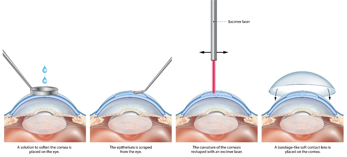
Photorefractive keratectomy (PRK)
The clear skin layer regrows over a period of about a week, then smooths off optically to complete the visual recovery over the next three months. While the skin layer is regrowing, the eye surface is normally very sore. This is one of the main differences between surface laser treatments and LASIK or SMILE, which both aim to keep the corneal skin layer intact. All surface laser treatments produce similar results, and the only difference between them is the way in which the corneal skin layer is removed. In PRK and LASEK the skin layer is removed by the surgeon – in LASEK dilute alcohol is applied to loosen the skin layer first. Some modern excimer laser systems are able to remove the skin layer as part of the reshaping treatment. This is called TransPRK. The area of skin layer removal in TransPRK is reduced to the minimum required for reshaping the cornea beneath, shortening recovery time by 1 to 2 days in comparison with PRK and LASEK.
Side effects are problems which most patients experience to some degree after surgery. They normally improve with time, but do not always resolve completely.

With modern laser systems, visual side effects are usually mild and improve within a few months. Lasting problems are unusual but may still occur.
Eye comfort
Other common side effects are intermittent blurring (variable vision) and eye surface discomfort (dry eye symptoms). Both are caused by reduced stability of the tear film between blinks. Tear film stability improves over a few months after treatment as the corneal surface heals. During the healing period, most patients are able to stay comfortable using tear supplements when required. Eye comfort usually returns to normal within a few months of treatment and, for contact lens wearers in particular, may be better after laser vision correction than before. For patients with a normal eye surface prior to surgery, lasting problems are unusual.
Eye Appearance
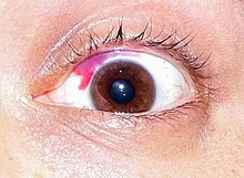
Red blotches are often visible on the white of the eye after any form of eye surgery, and are particularly common after LASIK. These are called subconjunctival haemorrhages and are caused by a small leak of blood under the clear membrane (the conjunctiva) covering the white part of eye wall. Although they can be quite unsightly, red blotches are temporary, and do not affect eye health; but they can take up to 6 weeks to go away completely.
If you develop a new eye health problem in later life, laser vision correction should not prevent you having successful treatment.
Common eye health problems like diabetic retinopathy and age-related macular degeneration are monitored and treated as normal after laser vision correction. Laser vision correction can affect eye pressure measurements used to check for glaucoma, causing them to under-read, especially in patients who have had treatment for high myopia. Corrections to eye pressure measurement can be applied to help ensure that glaucoma is picked up at an early stage and treated effectively; but it is worth reminding your optometrist or doctor that you have had laser vision correction when they are checking for or treating glaucoma. A record of your last prescription for glasses before laser vision correction may help them to make adjustments to your eye pressure readings more accurately. Laser vision correction can reduce the accuracy of focus correction and vision after future cataract surgery. Any detrimental effect is small and is reducing as more patients who have had previous laser vision correction are entering the cataract age group and modifications to lens implant selection calculations are becoming better understood. If you are having cataract surgery, remember to tell your surgeon that you have had laser vision correction. This helps to your surgeon to make the right modifications to lens implant selection.
You can eat and drink normally before surgery and should take any regular medication as usual. You have to be awake for laser vision correction so that you can look up to a target light. This helps you keep your eye in the right position during treatment. Stay as relaxed as you can during the surgery and try to keep your head still after the surgeon has positioned it comfortably for you. Most people are anxious prior to surgery. Your surgeon will be used to this, and will talk you through the procedure, encouraging you at every stage. Keep your breathing calm and tell your surgeon if you need a break. An anti-anxiety, muscle-relaxing drug such as diazepam can be helpful, particularly if you have a tendency to squeeze your eyes shut when they are being touched.
Discuss this with your surgeon before the day of surgery if you are worried. Modern laser systems either hold the eye still with gentle suction or have accurate tracking systems that follow eye movements during surgery, and a spring clip is used to hold the eyelids apart. So, you should not worry too much about moving or blinking during the procedure. But try to listen to instructions and keep your eyes on the fixation light when asked to do so. It is important to keep the eyes well lubricated in the first few hours after treatment, particularly after LASIK. Stay awake, but rest with your eyes closed when you can, and use lubricant drops frequently. You can wash and shower normally from day one after LASIK or SMILE, and once any bandage contact lenses used after surface laser treatment have been removed – typically on day 4 to day 7 after surgery. Most surgeons recommend no swimming for a week and, after LASIK, no contact sports for a month. Non-contact sports such as gym and jogging can be resumed from day one after surgery.
Set yourself a reminder and use the antibiotic and anti-inflammatory drops as prescribed to help the eyes to heal well. It is good to leave at least two minutes between different types of eye drop so that they each absorb well before the next drop is applied. If you miss the first time or you are not sure, applying a second eye drop is no problem. Some variability of vision and comfort is normal in the early weeks after surgery, and patience is required. But you should not be afraid to contact your surgeon if you have any concerns, or if you have an injury to the eye. You should contact your surgeon without delay if you have increasing pain, light sensitivity, redness, blur or an injury to the eye followed by pain, blur or watering. You may not be aware of a problem that requires treatment in the healing phase. So, make sure you attend your review appointments even if your eyes feel good.
Surgery to correct the need for glasses or contact lenses is not covered by private health insurance schemes. Your clinic should be clear from the outset about the total cost of the procedure. This normally includes follow up clinic visits and treatment for any problems resulting from surgery. Additional laser treatments to fine-tune the visual result are also normally included in this cost for up to two years after surgery. Although most prescription changes from three months after surgery are very small, it can take up to two years for the results of laser vision correction to stabilise fully. Most problems requiring further treatment would occur within this period. Most clinics do not accept an open-ended liability and will charge for additional treatment relating to natural prescription changes occurring later than two years after laser vision correction. Treatment or tests for any unrelated eye health problems are also normally charged separately.
Accommodation the reflex by which the eye brings a near object into focus by changing the shape of the natural lens. The natural lens gets less flexible with age, and powers of accommodation diminish. This affects reading vision and the ability to compensate for hyperopia.
Artisan/ArtiflexPIOL this is a type of PIOL that is clipped on to the iris during implantation. PIOLs are a commonly used alternative to laser vision correction for younger patients.
Astigmatism irregular defocus, or an eye that is ‘more rugby ball shaped than football shaped.’ The first number in your prescription for glasses describes the amount of long or short sight. The second number describes the amount of astigmatism. Most of us have at least some astigmatism, and a small amount can even help extend the range of activities you can do without reading glasses when you get older.
Biometry this test involves measurement of the eye and a set of calculations (biometry formulae) that help determine the right focusing power for an IOL before cataract surgery or RLE.
Binocular vision this is vision measured with the two eyes open.
Cataract when the natural lens gets misty enough to make vision hazy, it is called a cataract.
Cataract surgery this is surgery to replace the natural lens with an IOL. Cataract surgery is identical to RLE but is performed in patients who cannot see clearly despite using glasses or contact lenses with the main aim of restoring clear vision.
Conjunctiva the membrane covering the white of the eye. The conjunctiva produces mucus to help spread the watery tear film over the eye surface.
Cornea the clear part of the eye wall at the front of the eye. Two thirds of the focusing power of the eye is in the cornea.
Corneal epithelium the corneal skin layer
Corneal topography this is a scan that maps the surface curvature of the cornea.
D or Dioptre a unit for measuring the refractive power of a lens.
Excimer laser this type of laser removes tissue by non-thermal vaporisation (photoablation). Excimer lasers are extremely accurate and do not damage the surrounding tissues. They are used in LASIK and surface laser treatments (PRK, LASEK and TransPRK).
Femtosecond laser this type of laser is designed to cut any 3D shape in clear eye tissues such as the natural lens or the cornea with a high degree of accuracy. They work by creating a 3D pattern of tiny gas bubbles, which is traced through the target tissue at high speed. Femtosecond lasers are used in LASIK, SMILE, and increasingly in RLE and Cataract surgery.
Floaters floating shadows cast on the retina by opacities in the vitreous. Most of us are aware of floaters in some lighting conditions.
Glaucoma this is a condition in which the optic nerve is gradually damaged causing the visual field to contract. Left untreated, patients with glaucoma may develop ‘tunnel vision.’ Glaucoma is often associated with a higher than normal intraocular pressure, and treatment is centred on drugs or surgery to lower the intraocular pressure.
Hyperopia long sight. People with hyperopia typically have good vision as young adults. As they get older, they find themselves reliant on glasses for
reading, and then for the distance vision too. Younger people are able to compensate for hyperopia by accommodation. The amount of hyperopia is shown by a positive number in your spectacle prescription.
ICL (implantable contact lens) this is the most commonly used type of PIOL. It is implanted behind the iris and vaults over the natural lens – a bit like a contact lens implanted in the eye. ICL implantation is a commonly used alternative to laser vision correction in younger patients.
IOL (intraocular lens) IOLs are small synthetic lens implants that are used to replace the natural lens in cataract surgery and RLE.
Intraocular pressure (IOP) this is the pressure of fluid within the eye. It is often measured with a puff of air in routine eye checks or (more accurately) with a yellow drop and a blue light.
Iris this is the coloured part of the eye behind the cornea that expands and contracts in response to light to dilate or constrict the pupil.
LASEK (laser assisted sub-epithelial keratectomy) this is a form of surface laser treatment in which the corneal skin layer is soaked with dilute alcohol to loosen it before removal.
Laser vision correction correction of sight using excimer and/or femtosecond lasers to alter the curvature and focusing power of the cornea.
LASIK (laser in situ keratomileusis) this is the commonest form of laser vision correction in which a thin protective flap is created using a femtosecond laser. The protective flap is moved aside by the surgeon before optical reshaping of the cornea using an excimer laser. The flap is then replaced, and adheres without stitches, keeping the corneal skin layer intact and giving a fast visual recovery.
Meibomian glands the specialized oil glands in the eyelids that pump out a stabilizing layer of oil each time we blink that floats on top of the watery layer of the tear film. Inflammation of the eyelid margins upsetting this layer (blepharitis or meibomitis) is one of the commonest reasons for eye discomfort.
Micromonovision this is the name often given to the strategy of aiming for a clearer distance focus in one eye and a clearer focus at arms’ length in the other. Input from the two eyes combines to extend the range of focus for patients in the reading glasses age group undergoing vision correction surgery.
Monofocal IOL an IOL with one clear point of focus. These are the lenses most commonly used in standard cataract surgery. They have fewer optical side effects than multifocal lenses, but glasses are normally required for at least some activities after implantation.
Multifocal IOL an IOL with more than one point of clear focus This includes enhanced monofocal and Extended Depth of Focus lenses. Multifocal IOLs are often used in RLE in order to help increase freedom from glasses in the near range as well as providing good distance vision.
Myopia short sight. People with myopia are able to see up close but not in the distance. They typically first need glasses as school age children. The amount of myopia is shown as a negative number in your spectacle prescription.
Natural Lens the natural lens sits just behind the pupil and is suspended by a trampoline like array of microligaments from the ciliary muscle, which contracts during accommodation. The natural lens accounts for one third of the focusing power of the eye and is the flexible element of focus. The natural lens gets less flexible with age. It also becomes less clear as we get older. If the natural lens gets misty enough to make vision hazy, it is called a cataract.
Phacoemulsification this is the standard technique for liquefying the natural lens in cataract surgery and RLE. Energy delivered at ultrasonic frequencies from the tip of a fine, hollow probe liquefies the lens. Fluid is washed continuously into the eye around the probe, and the liquefied lens material is sucked away. Where previously the natural lens had to be shelled out like a pea, phacoemulsification allows it to be removed through a tiny, key-hole entry into the eye. This development revolutionised modern cataract surgery. Femtosecond lasers are now commonly used to break the lens up into small fragments before phacoemulsification.
Posterior capsule opacification (PCO) in cataract surgery and RLE, the IOL is implanted within the capsule of the natural lens. This thin, clear membrane then shrink-wraps the IOL and stabilises it in the natural position in the eye. As part of this healing up process, the membrane often goes misty, causing gradual loss of vision after surgery. This is posterior capsule opacification (PCO). It can be treated successfully with a one-off minor laser procedure called YAG capsulotomy.
Presbyopia age related loss of reading vision and the ability to focus on a near object without help from glasses.
PIOL (phakic intraocular lens) an eye that still has the natural lens in place is described as ‘phakic.’ PIOLs are small, synthetic lenses that are implanted in the eye without taking the natural lens out. PIOL implantation is an alternative to laser vision correction for younger patients.
Posterior vitreous detachment (PVD) as we age, the vitreous gel shrinks and will often peel off the back of the eye. For some of us, this event passes unnoticed. In others, a new shower of floaters will prompt them to attend for an eye examination. Doctors examining patients after a PVD look carefully for any abnormal attachment of the gel to the retina which could lead to a retinal detachment.
PRK (photorefractive keratectomy) this is a form of surface laser treatment in which preparatory removal of the corneal skin layer is done by the surgeon.
Refraction this is the test that is done to determine the numbers in your spectacle prescription and the amount of myopia, hyperopia or astigmatism.
Refractive surgery this is another name for vision correction surgery or surgery to reduce the need for glasses and contact lenses.
Retina the carpet of light sensitive cells lining the back of the eye, which is sometimes described as ‘the film in the camera.’ Images are focused on the retina by the cornea, the natural lens. Information from the retina is fed through the optic nerve to the visual areas of the brain.
Retinal detachment the retina sometimes detaches from the eye wall and its blood supply. Urgent surgery is then required to re-attach the retina in order to prevent visual loss. Patients with high myopia are more likely to get a retinal detachment. Retinal detachments are also more common in the early years after cataract surgery or RLE. Warning signs are a sudden change in vision with field loss (a dark shadow in part of the visual field); flashing lights (arcs of light – even with the eyes closed); and a sudden new shower of floaters.
RLE (refractive lens exchange) this is surgery to replace the natural lens with an IOL. RLE is identical to cataract surgery but is performed in patients who can see clearly if they wear glasses or contact lenses. The aim of RLE is to help people to see clearly for more activities without glasses or contact lenses.
SMILE (small incision lenticule extraction) this is a form of laser vision correction in which a lens shaped piece of corneal tissue is marked out using a femtosecond laser and removed surgically through a small incision.
Surface laser treatment a collective term for PRK, LASEK, TransPRK and other similar forms of laser vision correction in which optical reshaping of the cornea is performed on the corneal surface after removal of the corneal skin layer.
TransPRK (transepithelial PRK) this is a form of surface laser treatment in which the corneal skin layer is removed by the laser itself.
Tear film a multilayered wet film covering the front of the eye, which is essential for vision and comfort. A layer of mucus produced by specialized cells in the conjunctiva helps the watery layer of the tear film to spread over the eye. The watery layer is stabilized between blinks by an oily layer that forms a thin film (a bit like petrol floating on water) and acts to prevent localized evaporation. The oily layer is pumped out from specialized glands in the eyelids called meibomian glands each time we blink.
Visual acuity this is the main measurement of how well we can see and is tested by asking you to read down a chart which has smaller letters on each line. The lower you can read, the better your visual acuity is. Measurements are expressed as a fraction. If your visual acuity is 6/6 (normal) then you can see at 6 metres what a normally sighted person can see at 6 metres. If your vision is 6/9 you can see at 6 metres what a normally sighted person can see from 9 metres away et cetera. In the USA, the same measurement is made in feet, with 6 metres being roughly 20 feet. Many people are familiar with the American definition of normal ‘20/20’ vision. This is the same as 6/6 vision in the UK measurement.
Vitreous this is the gel filling the back of the eye. It tends to shrink as we age and accumulates wrinkles and opacities that cast floating shadows (floaters) on the retina.
Wavefront scan this is an optical map of your eye used to guide modern excimer laser treatments.
YAG capsulotomy a one-off minor laser procedure used to treat posterior capsule opacification (PCO).
Phakic Intraocular Lens Implantation
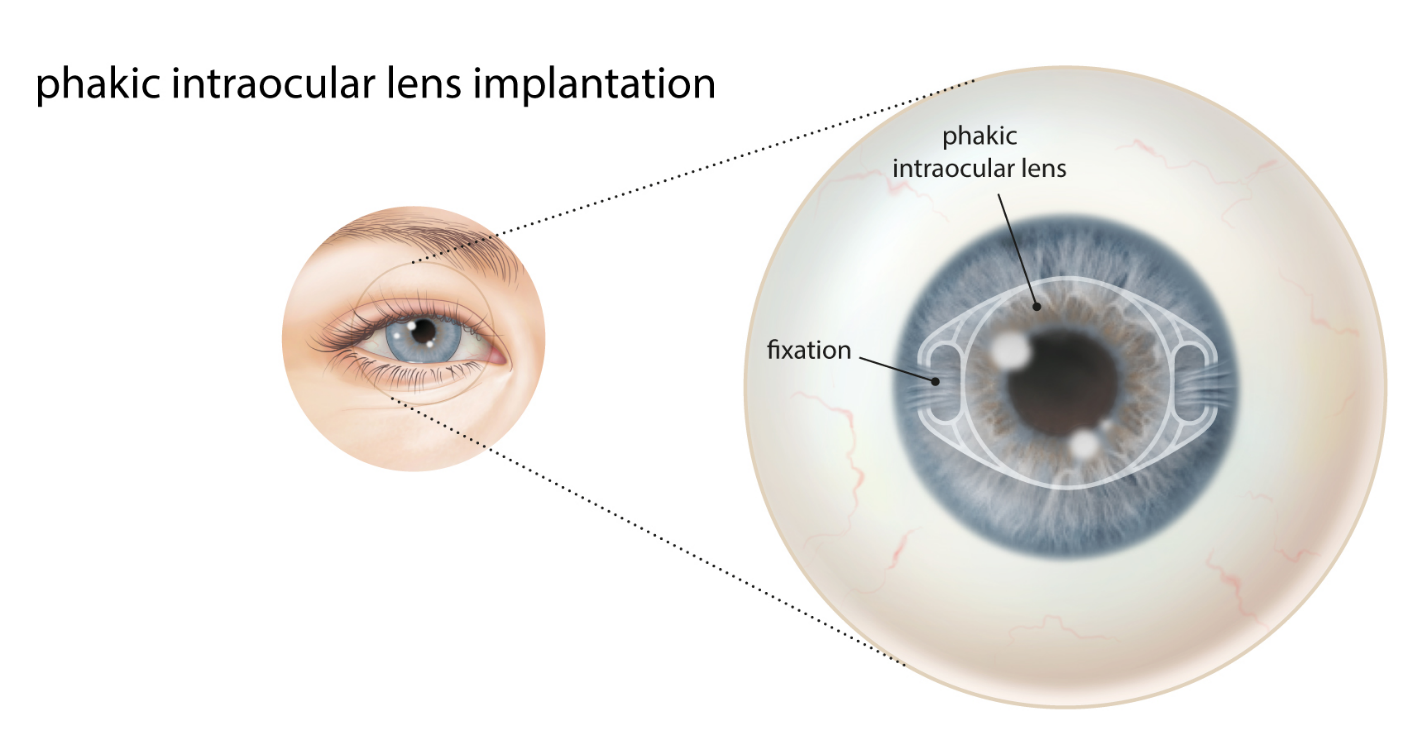
Lenses that are implanted into the eye to correct vision without taking out the natural lens are called phakic intraocular lenses (PIOLs). PIOLs are made of clear synthetic plastic. They sit either just in front of, or just behind, the pupil – a bit like building your contact lenses into your eyes. PIOL implantation is effective in treating high spectacle prescriptions and is widely used to treat younger patients who are not suitable for laser eye surgery.
The commonest type of PIOL implanted worldwide is the Visian ICL (implantable contact lens). This is a soft flexible implant designed to sit just behind the pupil and in front of the natural lens in the eye. You cannot see or feel ICLs after implantation and you do not need to clean them.
The other main type of PIOL is the Artisan/Artiflex PIOL. This PIOL clips onto the iris just in front of the pupil, and is sometimes visible as a glint in the eye. Like ICLs, you cannot feel Artisan/Artiflex PIOLs after implantation, and you do not need to clean them.
If you are suitable for PIOL implantation, your surgeon will discuss which type is the best option for you.
PIOLs are highly effective at treating both high glasses prescriptions and astigmatism.
For most patients, vision after PIOL surgery is similar to vision in contact lenses before surgery without the discomfort and limitations on activity.
Glasses may still be required for some activities after treatment, particularly reading in older patients, but these will be a low prescription and relatively inexpensive.
Over 95% of patients are satisfied with the outcome of surgery, and many describe it as life changing Although PIOL implantation is often bracketed with cosmetic surgery procedures, the benefits are mainly functional. It is designed to make you less dependent on glasses and contact lenses, helping you to lead an active lifestyle more easily. Short sight and astigmatism normally stabilize in the late teens or early 20s, but natural prescription changes can occur at any stage in life. So, laser vision correction is sometimes needed to enhance distance vision in the years after PIOL implantation.
Young patients who are unsuitable for laser vision correction are often offered PIOL implantation. This is because PIOLs can correct a wider range of spectacle prescriptions than laser vision correction and may be a safer option if you have pre-existing cornea or eye surface problems.
You need to be over 18 years of age and have a stable spectacle prescription. This is normally defined as no change greater than 0.5 units (0.5D) in the last 2 years.
The range of spectacle prescriptions that can be treated effectively is approximately:
- Up to -23.50D of myopia or short sight
- Up to +12.00D of hyperopia or long sight
- Up to ±4.50D of astigmatism
Supplementary laser vision correction can be used to extend this range for suitable patients with very high prescriptions in a combination treatment that is commonly called BIOPTICS. This is a combination treatment that includes lens and laser correction. You may not be suitable for PIOL implantation if you have other problems with your eye health including cataracts, glaucoma, or recurrent inflammation in the eye (uveitis). You also need to have enough room in the front of the eye to fit the PIOL safely. This is normally determined by a scan of the eye at your initial consultation.
Vision correction surgery alternatives
PIOL implantation is one of three main categories of operations to correct vision. The other two are laser vision correction and refractive lens exchange (RLE).
- Laser vision correction does not require a lens implant, and works by altering the curvature and focusing power of the front surface of the eye.
- RLE is identical to modern cataract surgery, and works by replacing the natural lens with a lens implant.
Laser vision correction is generally preferred if you have a lower glasses prescription. This is because there are very few longer-term risks associated with laser vision correction; whereas problems including cataract can occur many years after PIOL implantation. Older patients with a high spectacle prescription are more at risk of getting a cataract after PIOL implantation, and they have already lost most of the flexibility of focus provided by the natural lens in the eye. So after about 50 years of age, RLE is the usually the best option if you are unsuitable for laser vision correction. Your surgeon will advise on your best treatment options after reviewing your test measurements and your eye health.
Alternative PIOLs
Different types of PIOL have a different safety profile. Of the two main types now available (ICL and Artisan/ Artiflex), the ICL is easy to implant through a small entry into the eye and has a very good long-term safety record. Sizing can be an issue for the ICL, and some ICLs need to be replaced soon after the initial operation. Iris clip lenses (Artisan/Artiflex) have the advantage that one size fits all, but annual eye health monitoring is required after implantation.
Continuing in glasses or contact lenses
PIOL implantation is elective. This means you can choose to proceed with it at any time, or not at all. The alternative is staying in glasses or contact lenses Glasses are risk free but can be expensive for people with higher prescriptions and very limiting in terms of the range of activities, particularly sport, that you can participate in. Contact lenses provide good all-round vision. They do not mist over during sport and will help you to be more active; but they can be inconvenient when travelling, make water sports more difficult, and should not be worn whilst showering, swimming or during sleep. Contact lens wear is sometimes associated with eye surface discomfort and may be complicated by sight threatening infection. The risks and benefits of PIOL implantation should be balanced against those for continued contact lens wear, since this is the main alternative for active people considering vision correction surgery.
PIOL implantation is performed using eye-drop anaesthetic supplemented by an injection in the back of your hand to relax you if required. Anaesthetic may also be washed around the back of the eye to prevent excessive eye movement. A spring clip holding the eyelids apart allows you to blink safely during surgery.
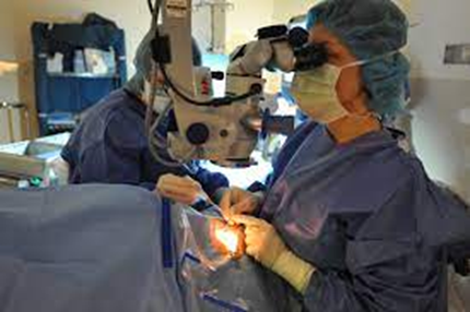
Your surgeon will be looking through a microscope to perform the surgery. You will be lying down under a surgical tent with fresh air coming in underneath. A sticky plastic drape covers the skin around the eye and sticks the eyelashes out of the way. Strong pupil dilating drugs are given as drops or a using a pellet placed under the lower eyelid to prepare the eye for surgery.
Essential steps in surgery are:
- Entry points – formation of small self-sealing entry points in the front of the eye at the junction of the white of the eye and the cornea
- PIOL insertion – injection of the PIOL and unfolding into position using a supporting gel to fill the front of the eye. Sometimes a small bypass drainage hole is formed in the iris at this point, but this additional stage is no longer required for newer (v4c and later) ICLs in myopic patients
- Wash out and refilling – washout of the supporting gel and refilling with fluid and antibiotics. Stitched closure of the eye is often required for Artisan PIOLs
Some centres offer surgery for both eyes on the same day. More commonly, second eye surgery is delayed for a day or longer to ensure that the recovery in the first eye is progressing well and, for ICLs, that sizing in the first eye is correct.
The surgery typically takes about 20 minutes per eye. You can return home on the same day as surgery.
In all forms of eye surgery, problems can occur during the operation or afterwards in the healing period. Problems can result in permanent, serious loss of vision (vision worse than the driving standard in the affected eye that cannot be corrected with glasses or contact lenses). More commonly, problems can be corrected with changes in medication or additional surgery. Typically, these additional operations feel like the original surgery and have a similar recovery period. Different types of PIOL have different associated risks. Your surgeon will ensure that you are given clear advice relevant to the lens type that is recommended.
Loss of vision
Permanent, serious loss of vision is very uncommon after ICL implantation. Causes include damage to the nerve at the back of the eye caused by a sudden rise in fluid pressure within the eye after surgery, and damage to the retina caused by infection or retinal detachment. Sudden pressure rises are much less common with the newer v4c ICL used for treating myopic patients. The v4c ICL allows natural fluid flow through the pupil and does not require a bypass drainage hole in the iris. If pressure rises do still occur, it is normally because of incomplete removal of supporting gel – a problem that can be fixed relatively easily with further washout. All patients with high levels of short sight have a higher risk of retinal detachment. This risk is not increased by ICL implantation, which does not involve surgery to the back compartment of the eye. Infection rates after ICL implantation are very low (approximately 1 in 6000). Complete loss of vision can occur after any operation involving the inside of the eye; but this is rare after ICL implantation. Although fluid pressure rises and infection can occur after Artisan/Artiflex PIOL implantation, the risk of problems leading to visual loss generally relates to later complications, particularly corneal clouding. These problems can often be spotted at an early stage and may be partially or completely intercepted by PIOL removal. Annual review with your eye surgeon for life is normally recommended after Artisan/Artiflex PIOL implantation, whereas a standard optometric eye health check once a year is sufficient after ICL implantation for which the main long-term risk is cataract formation. There is usually a charge for the annual review appointment.
Additional surgery
Cataracts may occur earlier in life than they would have done otherwise after all types of PIOL implantation. Cataract surgery can normally be combined with PIOL removal if necessary, and substitution of a new lens implant during cataract surgery helps to minimize any additional requirement for glasses. In other words, PIOL implantation does not stop you having successful cataract surgery or RLE later in life if this is required. Statistical techniques are used to size ICL implants. The size prediction is sometimes incorrect, and in approximately 1 case in 40, the ICL needs to be replaced with a lens of a different size in order to get the best fit in the eye. A minor rotation of the position of an ICL implant is also sometimes required after surgery to optimise the correction of astigmatism. For Artisan/Artiflex PIOLs, sizing is not a problem, since once size fits all. But repositioning procedures are sometimes required after the initial implantation. PIOLs can be removed if they are causing problems. This usually means that your vision and eye health will be the same as it was before PIOL implantation. But not all problems caused by PIOLs can be corrected by removing them, and additional treatment may be required even after PIOL removal.
ICL removal is uncommon unless you require cataract surgery or an ICL of a different size. Artisan/Artiflex PIOLs are sometimes removed to prevent further deterioration in the eye health if it looks like the risk of corneal clouding is increasing (as measured by annual counts of the endothelial cells on the back of the cornea as part of the annual visit), or if there are persistent problems with eye inflammation.
Risks of contact lens wear
Continuing in contact lenses is often the main alternative for people considering sight correction surgery. If you follow the right safety advice, contact lens wear is low risk; but approximately 1 in 3000 wearers each year will develop a serious corneal infection. To minimize this risk, you should not swim or shower in contact lenses, and should not wash them in tap water. Sleeping in contact lenses, including those designed for overnight wear, increases the risk of infection significantly. Soft, daily disposable lenses are safer than non-disposable lenses.
Side effects are problems which most patients experience to some degree after surgery. They normally improve with time, but do not always resolve completely.
Vision
It is normal to experience some light scatter side effects in the early period after PIOL implantation, particularly if you have treatment for a very high spectacle prescription. These can take a variety of forms including glare, halos, starbursts and ghost images. Increased flare from oncoming car headlights is a common symptom, and night driving may be difficult at first.
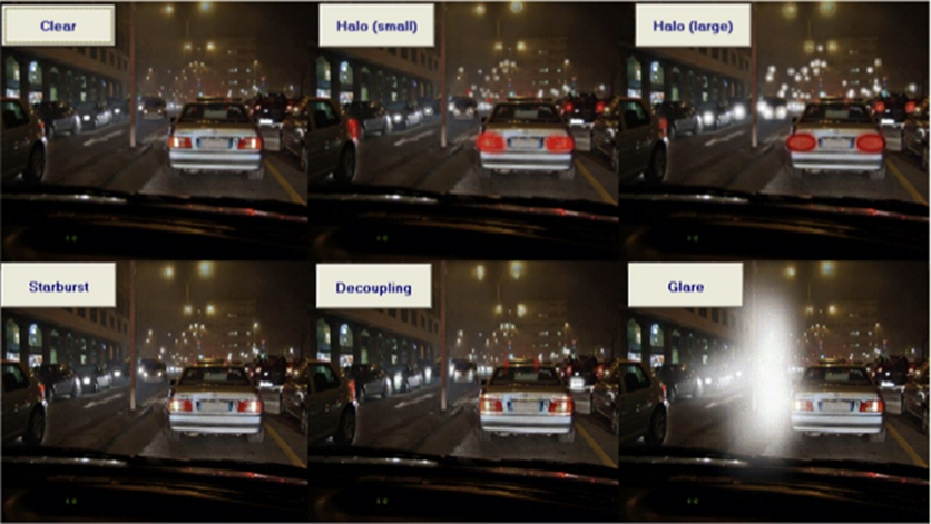
Visual side effects are usually mild and improve within a few months PIOLs can be removed if visual side effects persist, but this is rarely required.
Eye comfort
Some eye surface discomfort is common in the early months after most forms of eye surgery. This is usually mild after PIOL implantation, and highly variable – often affecting one eye more than the other. Treatment and prevention are based on making sure your eye surface is healthy before and after surgery. Lubricant eye drops can be helpful, and can be taken safely in addition to your other medication when required. For patients with a normal eye surface prior to surgery, lasting problems are unusual.
Eye appearance
Red blotches are often visible on the white of the eye after any form of eye surgery. These are called subconjunctival haemorrhages and are caused by a small leak of blood under the clear membrane covering the white part of eye wall. Although they can be quite unsightly, red blotches are temporary, and do not affect eye health; but they can take up to six weeks to go away completely.
ICLs are not visible. But you may be aware of a glint in the eye caused by a reflection from the lens after Artisan/Verisyse PIOL implantation.
If you develop a new eye health problem in later life, PIOL implantation should not prevent you having successful treatment. Common eye health problems like glaucoma, diabetic retinopathy, and age related macular degeneration can be monitored and treated as normal after PIOL implantation. If cataract surgery is required after PIOL implantation, the PIOL will have to be removed as part of the cataract procedure. ICLs can be removed safely through a normal cataract entry incision, whereas Artisan PIOL removal may require a larger entry. Some of the advantages of keyhole cataract surgery (no stitches, rapid recovery and less need for glasses) are reduced if a larger entry into the eye is required.
Most patients have PIOL implantation under local anaesthetic. You can eat and drink normally before surgery, and should take any regular medication as usual. Most surgeons work with an anaesthetist to monitor your health and optimise your comfort during surgery, administering sedation where necessary. Keep your breathing calm, stay as relaxed as you can, and try to keep your head still after the surgeon has positioned it comfortably. You can help your surgeon apply the drape and stick your eyelashes out of the way by opening both your eyes wide at the beginning of surgery. Blinking is no problem after the draping is complete. Just look straight up ahead to the bright operating light with both eyes open, but blink when you need to. Looking up to the bright microscope light helps to keep your eyes in the best position. Your surgeon will talk you through the procedure, encouraging you at every stage. Let your surgeon know if you feel any discomfort, and tell your surgeon if you need to cough, sneeze or take a break. A clear plastic shield may be taped over your eye at the end of surgery for protection on the way home.
Nursing staff will provide aftercare information and show you how to wear the eye shield at night (normally for 1 week after PIOL implantation). You can wash and shower normally from day one after surgery. Most surgeons recommend no swimming for a week and no contact sports for a month. Non-contact sports such as gym and jogging can be resumed from day one after surgery. Your surgeon will advise you when it is safe to start driving again. Typically, this is within a few days of surgery.
Set a reminder and use the antibiotic and anti- inflammatory drops as prescribed to help the eyes to heal well. It is good to leave at least two minutes between different types of eye drop so that they each absorb well before the next drop is applied. If you miss the first time or you are not sure, applying a second eye drop is no problem.
Some variability of vision and comfort is normal in the early weeks after PIOL implantation, and patience is required. But discomfort is usually mild, and vision normally recovers substantially within two to three days once the pupil dilating drugs have worn off. Report to your surgeon or an eye casualty department without delay if you have increasing aching pain, light sensitivity, redness, blur after surgery. You may not be aware of a problem that requires treatment in the healing phase. So, make sure you attend your review appointments even if your eyes feel good.
Surgery to correct the need for glasses or contact lenses is not covered by private health insurance schemes. Your clinic should be clear from the outset about the total cost of the procedure. Follow up clinic visits and treatment for any problems resulting from surgery are usually included in this cost for up to 12 months after surgery. Vision stabilizes quickly after PIOL implantation, but problems resulting from PIOL implantation, cataract in particular, may occur many years later. Most clinics do not accept an open-ended liability and will charge for additional treatment relating to natural prescription changes or a new problem with eye health.
Accommodation the reflex by which the eye brings a near object into focus by changing the shape of the natural lens. The natural lens gets less flexible with age, and powers of accommodation diminish. This affects reading vision and the ability to compensate for hyperopia.
Artisan/ArtiflexPIOL this is a type of PIOL that is clipped on to the iris during implantation. PIOLs are a commonly used alternative to laser vision correction for younger patients.
Astigmatism irregular defocus, or an eye that is ‘more rugby ball shaped than football shaped.’ The first number in your prescription for glasses describes the amount of long or short sight. The second number describes the amount of astigmatism. Most of us have at least some astigmatism, and a small amount can even help extend the range of activities you can do without reading glasses when you get older.
Biometry this test involves measurement of the eye and a set of calculations (biometry formulae) that help determine the right focusing power for an IOL before cataract surgery or RLE.
Binocular vision this is vision measured with the two eyes open.
Cataract when the natural lens gets misty enough to make vision hazy, it is called a cataract.
Cataract surgery this is surgery to replace the natural lens with an IOL. Cataract surgery is identical to RLE but is performed in patients who cannot see clearly despite using glasses or contact lenses with the main aim of restoring clear vision.
Conjunctiva the membrane covering the white of the eye. The conjunctiva produces mucus to help spread the watery tear film over the eye surface.
Cornea the clear part of the eye wall at the front of the eye. Two thirds of the focusing power of the eye is in the cornea.
Corneal epithelium the corneal skin layer
Corneal topography this is a scan that maps the surface curvature of the cornea.
D or Dioptre a unit for measuring the refractive power of a lens.
Excimer laser this type of laser removes tissue by non-thermal vaporisation (photoablation). Excimer lasers are extremely accurate and do not damage the surrounding tissues. They are used in LASIK and surface laser treatments (PRK, LASEK and TransPRK).
Femtosecond laser this type of laser is designed to cut any 3D shape in clear eye tissues such as the natural lens or the cornea with a high degree of accuracy. They work by creating a 3D pattern of tiny gas bubbles, which is traced through the target tissue at high speed. Femtosecond lasers are used in LASIK, SMILE, and increasingly in RLE and Cataract surgery.
Floaters floating shadows cast on the retina by opacities in the vitreous. Most of us are aware of floaters in some lighting conditions.
Glaucoma this is a condition in which the optic nerve is gradually damaged causing the visual field to contract. Left untreated, patients with glaucoma may develop ‘tunnel vision.’ Glaucoma is often associated with a higher than normal intraocular pressure, and treatment is centred on drugs or surgery to lower the intraocular pressure.
Hyperopia long sight. People with hyperopia typically have good vision as young adults. As they get older, they find themselves reliant on glasses for
reading, and then for the distance vision too. Younger people are able to compensate for hyperopia by accommodation. The amount of hyperopia is shown by a positive number in your spectacle prescription.
ICL (implantable contact lens) this is the most commonly used type of PIOL. It is implanted behind the iris and vaults over the natural lens – a bit like a contact lens implanted in the eye. ICL implantation is a commonly used alternative to laser vision correction in younger patients.
IOL (intraocular lens) IOLs are small synthetic lens implants that are used to replace the natural lens in cataract surgery and RLE.
Intraocular pressure (IOP) this is the pressure of fluid within the eye. It is often measured with a puff of air in routine eye checks or (more accurately) with a yellow drop and a blue light.
Iris this is the coloured part of the eye behind the cornea that expands and contracts in response to light to dilate or constrict the pupil.
LASEK (laser assisted sub-epithelial keratectomy) this is a form of surface laser treatment in which the corneal skin layer is soaked with dilute alcohol to loosen it before removal.
Laser vision correction correction of sight using excimer and/or femtosecond lasers to alter the curvature and focusing power of the cornea.
LASIK (laser in situ keratomileusis) this is the commonest form of laser vision correction in which a thin protective flap is created using a femtosecond laser. The protective flap is moved aside by the surgeon before optical reshaping of the cornea using an excimer laser. The flap is then replaced, and adheres without stitches, keeping the corneal skin layer intact and giving a fast visual recovery.
Meibomian glands the specialized oil glands in the eyelids that pump out a stabilizing layer of oil each time we blink that floats on top of the watery layer of the tear film. Inflammation of the eyelid margins upsetting this layer (blepharitis or meibomitis) is one of the commonest reasons for eye discomfort.
Micromonovision this is the name often given to the strategy of aiming for a clearer distance focus in one eye and a clearer focus at arms’ length in the other. Input from the two eyes combines to extend the range of focus for patients in the reading glasses age group undergoing vision correction surgery.
Monofocal IOL an IOL with one clear point of focus. These are the lenses most commonly used in standard cataract surgery. They have fewer optical side effects than multifocal lenses, but glasses are normally required for at least some activities after implantation.
Multifocal IOL an IOL with more than one point of clear focus This includes enhanced monofocal and Extended Depth of Focus lenses. Multifocal IOLs are often used in RLE in order to help increase freedom from glasses in the near range as well as providing good distance vision.
Myopia short sight. People with myopia are able to see up close but not in the distance. They typically first need glasses as school age children. The amount of myopia is shown as a negative number in your spectacle prescription.
Natural Lens the natural lens sits just behind the pupil and is suspended by a trampoline like array of microligaments from the ciliary muscle, which contracts during accommodation. The natural lens accounts for one third of the focusing power of the eye and is the flexible element of focus. The natural lens gets less flexible with age. It also becomes less clear as we get older. If the natural lens gets misty enough to make vision hazy, it is called a cataract.
Phacoemulsification this is the standard technique for liquefying the natural lens in cataract surgery and RLE. Energy delivered at ultrasonic frequencies from the tip of a fine, hollow probe liquefies the lens. Fluid is washed continuously into the eye around the probe, and the liquefied lens material is sucked away. Where previously the natural lens had to be shelled out like a pea, phacoemulsification allows it to be removed through a tiny, key-hole entry into the eye. This development revolutionised modern cataract surgery. Femtosecond lasers are now commonly used to break the lens up into small fragments before phacoemulsification.
Posterior capsule opacification (PCO) in cataract surgery and RLE, the IOL is implanted within the capsule of the natural lens. This thin, clear membrane then shrink-wraps the IOL and stabilises it in the natural position in the eye. As part of this healing up process, the membrane often goes misty, causing gradual loss of vision after surgery. This is posterior capsule opacification (PCO). It can be treated successfully with a one-off minor laser procedure called YAG capsulotomy.
Presbyopia age related loss of reading vision and the ability to focus on a near object without help from glasses.
PIOL (phakic intraocular lens) an eye that still has the natural lens in place is described as ‘phakic.’ PIOLs are small, synthetic lenses that are implanted in the eye without taking the natural lens out. PIOL implantation is an alternative to laser vision correction for younger patients.
Posterior vitreous detachment (PVD) as we age, the vitreous gel shrinks and will often peel off the back of the eye. For some of us, this event passes unnoticed. In others, a new shower of floaters will prompt them to attend for an eye examination. Doctors examining patients after a PVD look carefully for any abnormal attachment of the gel to the retina which could lead to a retinal detachment.
PRK (photorefractive keratectomy) this is a form of surface laser treatment in which preparatory removal of the corneal skin layer is done by the surgeon.
Refraction this is the test that is done to determine the numbers in your spectacle prescription and the amount of myopia, hyperopia or astigmatism.
Refractive surgery this is another name for vision correction surgery or surgery to reduce the need for glasses and contact lenses.
Retina the carpet of light sensitive cells lining the back of the eye, which is sometimes described as ‘the film in the camera.’ Images are focused on the retina by the cornea, the natural lens. Information from the retina is fed through the optic nerve to the visual areas of the brain.
Retinal detachment the retina sometimes detaches from the eye wall and its blood supply. Urgent surgery is then required to re-attach the retina in order to prevent visual loss. Patients with high myopia are more likely to get a retinal detachment. Retinal detachments are also more common in the early years after cataract surgery or RLE. Warning signs are a sudden change in vision with field loss (a dark shadow in part of the visual field); flashing lights (arcs of light – even with the eyes closed); and a sudden new shower of floaters.
RLE (refractive lens exchange) this is surgery to replace the natural lens with an IOL. RLE is identical to cataract surgery but is performed in patients who can see clearly if they wear glasses or contact lenses. The aim of RLE is to help people to see clearly for more activities without glasses or contact lenses.
SMILE (small incision lenticule extraction) this is a form of laser vision correction in which a lens shaped piece of corneal tissue is marked out using a femtosecond laser and removed surgically through a small incision.
Surface laser treatment a collective term for PRK, LASEK, TransPRK and other similar forms of laser vision correction in which optical reshaping of the cornea is performed on the corneal surface after removal of the corneal skin layer.
TransPRK (transepithelial PRK) this is a form of surface laser treatment in which the corneal skin layer is removed by the laser itself.
Tear film a multilayered wet film covering the front of the eye, which is essential for vision and comfort. A layer of mucus produced by specialized cells in the conjunctiva helps the watery layer of the tear film to spread over the eye. The watery layer is stabilized between blinks by an oily layer that forms a thin film (a bit like petrol floating on water) and acts to prevent localized evaporation. The oily layer is pumped out from specialized glands in the eyelids called meibomian glands each time we blink.
Visual acuity this is the main measurement of how well we can see and is tested by asking you to read down a chart which has smaller letters on each line. The lower you can read, the better your visual acuity is. Measurements are expressed as a fraction. If your visual acuity is 6/6 (normal) then you can see at 6 metres what a normally sighted person can see at 6 metres. If your vision is 6/9 you can see at 6 metres what a normally sighted person can see from 9 metres away et cetera. In the USA, the same measurement is made in feet, with 6 metres being roughly 20 feet. Many people are familiar with the American definition of normal ‘20/20’ vision. This is the same as 6/6 vision in the UK measurement.
Vitreous this is the gel filling the back of the eye. It tends to shrink as we age and accumulates wrinkles and opacities that cast floating shadows (floaters) on the retina.
Wavefront scan this is an optical map of your eye used to guide modern excimer laser treatments.
YAG capsulotomy a one-off minor laser procedure used to treat posterior capsule opacification (PCO).
Refractive Lens Exchange
Refractive Lens Exchange (RLE) is identical to modern cataract surgery. Both operations involve replacing the natural lens with an intraocular lens (IOL). The only difference is that cataract surgery is performed mainly to correct blur or light scatter caused by a misty natural lens; whereas RLE is performed to reduce the need for glasses or contact lenses. The focusing power of the IOL can be chosen to reduce dependence on glasses after surgery.
IOL implantation is a bit like building your glasses or contact lenses into your eyes.
There are three main IOL types:
- Monofocal IOLs – monofocal IOLs aim to reduce spectacle dependence for distance (driving) vision
- Multifocal IOLs – multifocal IOLs aim to reduce spectacle dependence for a wider range of activities, including intermediate (computer screens) and near (reading) vision
- Extended depth of focus (EDOF) and enhanced monofocal IOLs – provide good distance and intermediate vision without glasses but reading glasses may be required. Reading vision may be proved without glasses by undercorrecting one eye for distance to make it slightly short-sighted.
If you are suitable for RLE, your surgeon will discuss which IOL type is the best option for you.
About four out of five patients are completely free of glasses after RLE with widely used contemporary multifocal IOLs.
If you do need help from glasses after surgery, it is usually for a specific activity in the near, intermediate or distance range.
Approximately 95% of patients are satisfied with the outcome of surgery, and many describe it as life changing.
Although RLE is often bracketed with cosmetic surgery procedures, the benefits are mainly functional. It is designed to make you less dependent on glasses and contact lenses, helping you to lead an active lifestyle more easily. RLE eliminates the need for cataract surgery in later life, and it is often preferred to laser vision correction for patients in the retirement age group in whom the early stages of cataract formation are often already present. In the absence of a new health problem in the eye, vision normally remains good, and your spectacle prescription normally remains stable after RLE. If you do experience any deterioration in vision in the years after surgery, it can often be reversed with a one-off minor laser procedure called YAG capsulotomy.
If you are over 50 years of age and have a glasses prescription you are likely to be suitable for RLE.
Almost any level of myopia (short sight) or hyperopia (long sight) can be corrected by RLE, and IOLs with built in astigmatism correction are available. Lens implantation techniques can be combined with laser eye surgery in suitable patients to extend the range of astigmatism treatment or fine-tune the focus outcome.
You may not be suitable for multifocal IOL implantation if you have pre-existing problems with your eye health.
Vision correction surgery alternatives
RLE is one of three main categories of operations to correct vision. The other two are laser vision correction and phakic intraocular lens (PIOL) implantation.
- Laser vision correction does not require a lens implant, and works by altering the curvature and focusing power of the front surface of the eye
- PIOLs are lens implants that sit in front of the natural lens rather than replacing it
Laser eye surgery or PIOL implantation are generally better options than RLE for younger patients who still have a clear, flexible natural lens. This includes most patients under 50 years old. Laser vision correction is a relatively low risk option for many patients over 50 years of age with a lower prescription; but the balance shifts as you get older and both flexibility and clarity of the natural lens diminish. RLE is the default option for vision correction surgery in the over 65 age group, but laser vision correction may still be a better alternative for patients with no signs of cataract and good eye surface health. Your surgeon will advise on your best treatment options after reviewing your test measurements and your eye health.
IOL alternatives
Different lens implants suit different patients, and not everyone will benefit from multifocal or EDOF IOL implantation. Monofocal IOLs may be more suitable if you have other eye problems or if you are worried that you may not cope well with the optical side effects of multifocal IOL implantation. Monofocal IOLs have fewer optical side effects than multifocal IOLs, but do not provide freedom from glasses for such a wide range of activities. The recently introduced enhanced monofocals offer a greater range of vision than standard monofocals with good intermediate vision. They do not have the same issues as multifocal IOLs in terms of unwanted visual disturbances. The most commonly used approach in standard cataract surgery is to select monofocal IOLs aiming to leave your eyes equally focused in the distance. Glasses are then usually required for reading and near work. A common alternative approach using monofocal IOLs is to aim for clearer distance vision in one eye, and clearer vision at arms’ length in the other. This works very well with enhanced monofocal IOLs to give a greater degree of spectacle freedom. With both eyes open, binocular visual input combines to extend the range of focus, improving vision for computer screens and working with your hands. Although with standard monofocals you would normally still need glasses for reading, near vision is at least partly restored, with relatively little compromise optically. This spread of binocular focus is sometimes called micromonovision. This works very well with enhanced monofocal IOLs. These can be a good intermediate choice if you would like more freedom from glasses but do not want, or do not have access to, multifocal IOLs. Your surgeon will discuss the pros and cons of different implant choices in RLE with you at your initial consultation.
Continuing in glasses or contact lenses.
RLE is elective. This means you can choose to proceed with it at any time, or not at all. The alternative is staying in glasses or contact lenses. Glasses are risk free but may limit the range of activities you can do confidently and comfortably – particularly sport and exercise. Although there are many successful lifelong contact lens wearers, comfort and tolerance tends to diminish with age. Varifocal glasses are often the main alternative to RLE for older patients. Varifocal glasses have their own optical side effects. For example, having the lower part of the varifocal lens focused for reading can make it difficult to walk downstairs confidently. Multifocal IOLs provide the range of focus in a different way, and trouble adapting to varifocal glasses does not mean that you will also have trouble adapting to multifocal IOLs.
Contact lenses provide good all-round vision. They do not mist over during sport and will help you to be more active; but they can be inconvenient when travelling, make water sports more difficult, and should not be worn whilst showering or during sleep. Contact lens wear is sometimes associated with eye surface discomfort, and may be complicated by sight threatening infection.
Risks, side effects and benefits of RLE should be balanced against those for continued contact lens wear and varifocal glasses since these are the main alternatives for people considering sight correction surgery.
RLE is performed using drop or gel anaesthetic supplemented by an injection in the back of your hand to relax you if required. Anaesthetic may also be washed around the back of the eye to prevent excessive eye movement. A spring clip holding the eyelids apart allows you to blink safely during surgery.
Your surgeon will be looking through a microscope to perform the surgery. You will be lying down under a surgical tent with fresh air coming in underneath. A sticky plastic drape covers the skin around your eye and sticks the eyelashes out of the way.
Many centres now offer surgery for both eyes on the same day, but you can still have surgery in one eye at a time if that is what you would prefer. Your surgeon will talk you through the pros and cons of same day surgery for both eyes at your consultation. The surgery typically takes about 20 minutes per eye. You can return home on the same day as surgery. Strong pupil dilating drugs are given as drops or as a pellet placed under the lower eyelid to prepare your eye for surgery. Essential steps in surgery are:
- Entry points – formation of small self-sealing entry points in the front of the eye at the junction of the white of the eye and the cornea
- Capsulotomy – removal of a circular disc from the front of the membrane covering the natural lens called the lens capsule (think of the skin of a grape and you will have the right mental image of the thin, clear covering of the natural lens)
- Phacoemulsification – liquefaction and removal of the natural lens from within the lens capsule using a high frequency vibrating probe, fluid washing, and vacuum IOL insertion – injection of the IOL and unfolding into the natural position within the lens capsule using a supporting gel to fill the front of the eye
- Wash out and refilling – wash-out of the supporting gel and refilling with fluid and antibiotics.
Femtosecond lasers are sometimes now used in a preparatory stage before going through to the operating theatre. Femtosecond lasers are costly and provide no proven benefit for your vision after RLE or cataract surgery, but they may make some stages of the operation (capsulotomy in particular) easier for surgeons to perform safely and consistently. This additional laser stage takes about five minutes and is performed using eye-drops to anaesthetise your eye.
In all forms of eye surgery, problems can occur during the operation or afterwards in the healing period. Problems can result in permanent, serious loss of vision (vision worse than the driving standard in the affected eye that cannot be corrected with glasses or contact lenses). More commonly, problems can be corrected with changes in medication or additional surgery. Typically, these additional operations feel like the original surgery and have a similar recovery period.
Loss of vision
Permanent, serious loss of vision is significantly more common after RLE than after laser vision correction or PIOL implantation, affecting approximately 1 in 500 patients. This may mean vision worse than the driving standard or, in some cases, complete loss of vision in the affected eye. Permanent serious visual loss is typically caused by damage to the retina. This can result from infection or an inflammatory response after surgery, retinal detachment after surgery, or bleeding during surgery. Some problems occurring during surgery increase the risk of sight threatening problems afterwards. These include a common complication of surgery called posterior capsular rupture, (breakage of the membrane just behind the IOL). One national benchmark rate for capsule rupture for all cataract surgeons is just under 1.5%. Experienced surgeons have a lower rate, but all surgeons have at least some cases affected by posterior capsular rupture. It is not always possible to implant a multifocal IOL safely if posterior capsular rupture has occurred, and this may mean greater than anticipated reliance on glasses after surgery.
Additional surgery
Second operations may be required to correct a complication from the initial surgery. This could include lens repositioning or exchange, surgery to retrieve lens fragments from the back of the eye, or retinal detachment repair. With or without RLE, retinal detachments are more common if you are very short-sighted; but the risk of retinal detachment is approximately five times higher in the first four years after cataract surgery or refractive lens exchange. Statistical techniques (biometry formulae) and eye measurements (biometry) are used to guide selection of the IOL required to correct your vision. Limitations on the accuracy of these techniques mean that laser vision correction to fine tune the focus is sometimes required to touch up the visual result after RLE. The commonest reason for visual deterioration after RLE is posterior capsule opacification (PCO). This is a gradual misting over of the membrane just behind the IOL, which affects many patients. PCO may occur months or years after surgery and is normally treated successfully with a one-off minor laser procedure called YAG laser capsulotomy.
Risks of contact lens wear
Continuing in contact lenses is often the main alternative for people considering sight correction surgery. If you follow the right safety advice, contact lens wear is low risk; but approximately 1 in 3000 wearers each year will develop a serious corneal infection. To minimise this risk, you should not swim or shower in contact lenses, and should not wash them in tap water. Sleeping in contact lenses, including those designed for overnight wear, increases the risk of infection significantly. Soft, daily disposable lenses are safer than non-disposable lenses.
Side effects are problems which most patients experience to some degree after surgery. They normally improve with time, but do not always resolve completely.
Vision
Most patients experience some light scatter side effects and unwanted images in the early months after RLE. Visual side effects vary with type of IOL implanted and are often more noticeable in some lighting conditions than others. Patients are commonly aware of a shadow or shimmering arc of light in their peripheral vision after monofocal or multifocal IOL implantation. Some types of multifocal IOLs tend to cause halos around lights. Blur or ghost images are more common with others; but almost any form of light scatter side effect can occur. It is generally accepted that all forms of multifocal IOL are associated with more optical side effects than monofocal IOLs. However, multifocal IOLs all produce a greater range of glasses freedom. Optical side effects may initially interfere with work or leisure activities, and night driving in particular. But they tend to diminish with time.
19 out of 20 patients are satisfied or very satisfied with their vision three to six-months after multifocal IOL implantation, and laser procedures to treat residual defocus or posterior capsular opacification (YAG capsulotomy for PCO) are often helpful in accelerating adaptation in the remainder. But approximately 1% of patients cannot adapt, and will elect to undergo IOL exchange, a potentially complicated procedure swapping the multifocal IOL for a monofocal IOL. The availability of enhanced monofocal IOLs provide an alternative at IOL exchange as their visual disturbances are much less than multifocal IOLs without complete loss of spectacle independence.
Eye comfort
Some eye surface discomfort is common in the early months after most forms of eye surgery. This is usually mild after RLE, and highly variable – often affecting one eye more than the other. Treatment and prevention are based on making sure your eye surface is healthy before and after surgery. Lubricant eye drops can be helpful and can be taken safely in addition to your other medication when required. For patients with a normal eye surface prior to surgery, lasting problems are unusual.
Eye Appearance
Red blotches are often visible on the white of the eye after any form of eye surgery. These are called subconjunctival haemorrhages and are caused by a small leak of blood under the clear membrane covering the white part of eye wall. Although they can be quite unsightly, red blotches are temporary, and do not affect eye health; but they can take up to six weeks to go away completely.

Most IOLs are not visible. But people may occasionally notice a glint in your eye caused by a reflection from the front of the IOL within the pupil.
If you develop a new eye health problem in later life, RLE implantation should not prevent you having successful treatment. Common eye health problems like glaucoma, diabetic retinopathy, and age- related macular degeneration can be monitored and treated as normal after RLE.
Many patients with hyperopia (long sight) have a relatively small space for fluid circulation through the front of the eye. The space narrows as the natural lens expands with age, leaving these patients vulnerable to a form of glaucoma caused by blockage of fluid flow that can lead to sudden, painful loss of sight (‘angle closure glaucoma’). In patients with this predisposition to later problems called ‘a shallow anterior chamber’, RLE can both reduce the need for glasses and remove any risk of future problems due to angle closure. This is because IOLs are thinner than the natural lens. So RLE or cataract surgery creates more space for fluid circulation in the front of the eye.
Most patients have IOL implantation under local anaesthetic. You can eat and drink normally before surgery, and should take any regular medication as usual.
Most surgeons work with an anaesthetist to monitor your health during surgery and optimise your comfort, administering sedation where necessary. Keep your breathing calm, stay as relaxed as you can, and try to keep your head still after the surgeon has positioned it comfortably. You can help your surgeon apply the drape and stick your eyelashes out of the way by opening both your eyes wide at the beginning of surgery. Blinking is no problem after the draping is complete. Just look straight up ahead to the bright operating light with both eyes open, but blink when you need to. Looking up to the bright microscope light helps to keep your eyes in the best position. Your surgeon will talk you through the procedure, encouraging you at every stage. Let your surgeon know if you feel any discomfort, and tell your surgeon if you need to cough, sneeze or take a break. For the preliminary laser stage in laser assisted RLE, your eye is normally held still and the eyelids are kept out of the way with a suction ring. But the same principles apply. To help the surgery go smoothly, simply stare straight up ahead with both eyes open. Try not to squeeze the eyes shut, but blink whenever you need to.
A clear plastic shield may be taped over the eye at the end of surgery to protect the eye on the way home. Nursing staff will show you how to wear the eye shield at night (normally for one week after RLE). You can wash and shower normally from day one after RLE. Most surgeons recommend no swimming for a week and no contact sports for a month. Non- contact sports such as gym and jogging can be resumed from day one after surgery. Your surgeon will advise you when it is safe to start driving again. Typically, this is within a few days of surgery. Set a smart phone reminder and use the antibiotic and anti-inflammatory drops as prescribed to help the eyes to heal well. It is good to leave at least two minutes between different types of eye drop so that they each absorb well before the next drop is applied. If you miss the first time or you are not sure, applying a second eye drop is no problem. Some variability of vision and comfort is normal in the early weeks after RLE, and patience is required. But discomfort is usually mild, and vision normally recovers substantially within two to three days once the pupil dilating drugs have worn off.
Report to your surgeon or an eye emergency department without delay if you have increasing aching pain, light sensitivity, redness, blur after surgery. Other danger signs, particularly relevant to highly myopic (short-sighted) patients undergoing RLE, relate to the warning signs of a retinal detachment. These are a sudden new shower of floaters, flashes of light (even with the eyes closed) and visual field loss (a shadow or curtain spreading across your vision). Most retinal detachments can be repaired without detriment to your vision, but the chances of success are much higher if the detachment has not spread across the centre of your vision. You may not be aware of a problem that requires treatment in the healing phase. So make sure you attend your review appointments even if your eyes feel good.
Surgery to correct the need for glasses or contact lenses is not covered by private health insurance schemes. Your clinic should be clear from the outset about the total cost of the procedure. Factors affecting cost include the type of IOL used and the use of femtosecond lasers. Monofocal IOLs are generally less expensive than multifocal and EDOF and enhanced monofocal IOLs, but IOL cost varies widely. Femtosecond lasers are now often used to cut the templates accurately for the initial stages of RLE. This expensive new technology may increase the cost of your procedure.
Follow up clinic visits and treatment for any problems resulting from surgery are usually included in the procedure cost for up to 12 months after surgery. Laser adjustments to focus are often required to obtain the best result from RLE. These are also normally included in the procedure fee. Most clinics do not accept an open-ended liability and will charge for additional treatment relating to natural prescription changes or a new problem with eye health. An additional fee may be charged for YAG capsulotomy, a one-off minor laser procedure that is often required months or years after RLE.
Accommodation the reflex by which the eye brings a near object into focus by changing the shape of the natural lens. The natural lens gets less flexible with age, and powers of accommodation diminish. This affects reading vision and the ability to compensate for hyperopia.
Artisan/ArtiflexPIOL this is a type of PIOL that is clipped on to the iris during implantation. PIOLs are a commonly used alternative to laser vision correction for younger patients.
Astigmatism irregular defocus, or an eye that is ‘more rugby ball shaped than football shaped.’ The first number in your prescription for glasses describes the amount of long or short sight. The second number describes the amount of astigmatism. Most of us have at least some astigmatism, and a small amount can even help extend the range of activities you can do without reading glasses when you get older.
Biometry this test involves measurement of the eye and a set of calculations (biometry formulae) that help determine the right focusing power for an IOL before cataract surgery or RLE.
Binocular vision this is vision measured with the two eyes open.
Cataract when the natural lens gets misty enough to make vision hazy, it is called a cataract.
Cataract surgery this is surgery to replace the natural lens with an IOL. Cataract surgery is identical to RLE but is performed in patients who cannot see clearly despite using glasses or contact lenses with the main aim of restoring clear vision.
Conjunctiva the membrane covering the white of the eye. The conjunctiva produces mucus to help spread the watery tear film over the eye surface.
Cornea the clear part of the eye wall at the front of the eye. Two thirds of the focusing power of the eye is in the cornea.
Corneal epithelium the corneal skin layer
Corneal topography this is a scan that maps the surface curvature of the cornea.
D or Dioptre a unit for measuring the refractive power of a lens.
Excimer laser this type of laser removes tissue by non-thermal vaporisation (photoablation). Excimer lasers are extremely accurate and do not damage the surrounding tissues. They are used in LASIK and surface laser treatments (PRK, LASEK and TransPRK).
Femtosecond laser this type of laser is designed to cut any 3D shape in clear eye tissues such as the natural lens or the cornea with a high degree of accuracy. They work by creating a 3D pattern of tiny gas bubbles, which is traced through the target tissue at high speed. Femtosecond lasers are used in LASIK, SMILE, and increasingly in RLE and Cataract surgery.
Floaters floating shadows cast on the retina by opacities in the vitreous. Most of us are aware of floaters in some lighting conditions.
Glaucoma this is a condition in which the optic nerve is gradually damaged causing the visual field to contract. Left untreated, patients with glaucoma may develop ‘tunnel vision.’ Glaucoma is often associated with a higher than normal intraocular pressure, and treatment is centred on drugs or surgery to lower the intraocular pressure.
Hyperopia long sight. People with hyperopia typically have good vision as young adults. As they get older, they find themselves reliant on glasses for
reading, and then for the distance vision too. Younger people are able to compensate for hyperopia by accommodation. The amount of hyperopia is shown by a positive number in your spectacle prescription.
ICL (implantable contact lens) this is the most commonly used type of PIOL. It is implanted behind the iris and vaults over the natural lens – a bit like a contact lens implanted in the eye. ICL implantation is a commonly used alternative to laser vision correction in younger patients.
IOL (intraocular lens) IOLs are small synthetic lens implants that are used to replace the natural lens in cataract surgery and RLE.
Intraocular pressure (IOP) this is the pressure of fluid within the eye. It is often measured with a puff of air in routine eye checks or (more accurately) with a yellow drop and a blue light.
Iris this is the coloured part of the eye behind the cornea that expands and contracts in response to light to dilate or constrict the pupil.
LASEK (laser assisted sub-epithelial keratectomy) this is a form of surface laser treatment in which the corneal skin layer is soaked with dilute alcohol to loosen it before removal.
Laser vision correction correction of sight using excimer and/or femtosecond lasers to alter the curvature and focusing power of the cornea.
LASIK (laser in situ keratomileusis) this is the commonest form of laser vision correction in which a thin protective flap is created using a femtosecond laser. The protective flap is moved aside by the surgeon before optical reshaping of the cornea using an excimer laser. The flap is then replaced, and adheres without stitches, keeping the corneal skin layer intact and giving a fast visual recovery.
Meibomian glands the specialized oil glands in the eyelids that pump out a stabilizing layer of oil each time we blink that floats on top of the watery layer of the tear film. Inflammation of the eyelid margins upsetting this layer (blepharitis or meibomitis) is one of the commonest reasons for eye discomfort.
Micromonovision this is the name often given to the strategy of aiming for a clearer distance focus in one eye and a clearer focus at arms’ length in the other. Input from the two eyes combines to extend the range of focus for patients in the reading glasses age group undergoing vision correction surgery.
Monofocal IOL an IOL with one clear point of focus. These are the lenses most commonly used in standard cataract surgery. They have fewer optical side effects than multifocal lenses, but glasses are normally required for at least some activities after implantation.
Multifocal IOL an IOL with more than one point of clear focus This includes enhanced monofocal and Extended Depth of Focus lenses. Multifocal IOLs are often used in RLE in order to help increase freedom from glasses in the near range as well as providing good distance vision.
Myopia short sight. People with myopia are able to see up close but not in the distance. They typically first need glasses as school age children. The amount of myopia is shown as a negative number in your spectacle prescription.
Natural Lens the natural lens sits just behind the pupil and is suspended by a trampoline like array of microligaments from the ciliary muscle, which contracts during accommodation. The natural lens accounts for one third of the focusing power of the eye and is the flexible element of focus. The natural lens gets less flexible with age. It also becomes less clear as we get older. If the natural lens gets misty enough to make vision hazy, it is called a cataract.
Phacoemulsification this is the standard technique for liquefying the natural lens in cataract surgery and RLE. Energy delivered at ultrasonic frequencies from the tip of a fine, hollow probe liquefies the lens. Fluid is washed continuously into the eye around the probe, and the liquefied lens material is sucked away. Where previously the natural lens had to be shelled out like a pea, phacoemulsification allows it to be removed through a tiny, key-hole entry into the eye. This development revolutionised modern cataract surgery. Femtosecond lasers are now commonly used to break the lens up into small fragments before phacoemulsification.
Posterior capsule opacification (PCO) in cataract surgery and RLE, the IOL is implanted within the capsule of the natural lens. This thin, clear membrane then shrink-wraps the IOL and stabilises it in the natural position in the eye. As part of this healing up process, the membrane often goes misty, causing gradual loss of vision after surgery. This is posterior capsule opacification (PCO). It can be treated successfully with a one-off minor laser procedure called YAG capsulotomy.
Presbyopia age related loss of reading vision and the ability to focus on a near object without help from glasses.
PIOL (phakic intraocular lens) an eye that still has the natural lens in place is described as ‘phakic.’ PIOLs are small, synthetic lenses that are implanted in the eye without taking the natural lens out. PIOL implantation is an alternative to laser vision correction for younger patients.
Posterior vitreous detachment (PVD) as we age, the vitreous gel shrinks and will often peel off the back of the eye. For some of us, this event passes unnoticed. In others, a new shower of floaters will prompt them to attend for an eye examination. Doctors examining patients after a PVD look carefully for any abnormal attachment of the gel to the retina which could lead to a retinal detachment.
PRK (photorefractive keratectomy) this is a form of surface laser treatment in which preparatory removal of the corneal skin layer is done by the surgeon.
Refraction this is the test that is done to determine the numbers in your spectacle prescription and the amount of myopia, hyperopia or astigmatism.
Refractive surgery this is another name for vision correction surgery or surgery to reduce the need for glasses and contact lenses.
Retina the carpet of light sensitive cells lining the back of the eye, which is sometimes described as ‘the film in the camera.’ Images are focused on the retina by the cornea, the natural lens. Information from the retina is fed through the optic nerve to the visual areas of the brain.
Retinal detachment the retina sometimes detaches from the eye wall and its blood supply. Urgent surgery is then required to re-attach the retina in order to prevent visual loss. Patients with high myopia are more likely to get a retinal detachment. Retinal detachments are also more common in the early years after cataract surgery or RLE. Warning signs are a sudden change in vision with field loss (a dark shadow in part of the visual field); flashing lights (arcs of light – even with the eyes closed); and a sudden new shower of floaters.
RLE (refractive lens exchange) this is surgery to replace the natural lens with an IOL. RLE is identical to cataract surgery but is performed in patients who can see clearly if they wear glasses or contact lenses. The aim of RLE is to help people to see clearly for more activities without glasses or contact lenses.
SMILE (small incision lenticule extraction) this is a form of laser vision correction in which a lens shaped piece of corneal tissue is marked out using a femtosecond laser and removed surgically through a small incision.
Surface laser treatment a collective term for PRK, LASEK, TransPRK and other similar forms of laser vision correction in which optical reshaping of the cornea is performed on the corneal surface after removal of the corneal skin layer.
TransPRK (transepithelial PRK) this is a form of surface laser treatment in which the corneal skin layer is removed by the laser itself.
Tear film a multilayered wet film covering the front of the eye, which is essential for vision and comfort. A layer of mucus produced by specialized cells in the conjunctiva helps the watery layer of the tear film to spread over the eye. The watery layer is stabilized between blinks by an oily layer that forms a thin film (a bit like petrol floating on water) and acts to prevent localized evaporation. The oily layer is pumped out from specialized glands in the eyelids called meibomian glands each time we blink.
Visual acuity this is the main measurement of how well we can see and is tested by asking you to read down a chart which has smaller letters on each line. The lower you can read, the better your visual acuity is. Measurements are expressed as a fraction. If your visual acuity is 6/6 (normal) then you can see at 6 metres what a normally sighted person can see at 6 metres. If your vision is 6/9 you can see at 6 metres what a normally sighted person can see from 9 metres away et cetera. In the USA, the same measurement is made in feet, with 6 metres being roughly 20 feet. Many people are familiar with the American definition of normal ‘20/20’ vision. This is the same as 6/6 vision in the UK measurement.
Vitreous this is the gel filling the back of the eye. It tends to shrink as we age and accumulates wrinkles and opacities that cast floating shadows (floaters) on the retina.
Wavefront scan this is an optical map of your eye used to guide modern excimer laser treatments.
YAG capsulotomy a one-off minor laser procedure used to treat posterior capsule opacification (PCO).